3D Curved Multiplanar Reformatting Provides Improved Visualization of Hippocampal Anatomy
Abstract number :
2.173
Submission category :
5. Neuro Imaging / 5A. Structural Imaging
Year :
2018
Submission ID :
501467
Source :
www.aesnet.org
Presentation date :
12/2/2018 4:04:48 PM
Published date :
Nov 5, 2018, 18:00 PM
Authors :
Ehsan Misaghi, University of Alberta; Trevor A. Steve, University of Alberta; Alan Wilman, University of Alberta; Christian Beaulieu, University of Alberta; and Donald W. Gross, University of Alberta
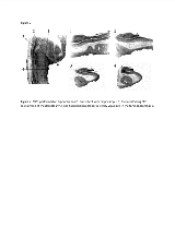
Rationale: Hippocampal Sclerosis (HS) is known to demonstrate considerable inter-subject heterogeneity regarding pathological involvement of different hippocampal subfields[1] as well as variable findings along the caudal – rostral axis of the hippocampus [2]. While there have been tremendous advances in the evaluation of hippocampal subfields with in vivo MRI, most protocols have been restricted to the hippocampal body where the classical interlocking “C” relationship of the hippocampus proper and dentate gyrus are readily visualized in the coronal plane. The anatomical relationship of the dentate gyrus and hippocampus proper is much more difficult to visualize in the caudal hippocampus (head), and the reliability of segmentation protocols for the hippocampal head has been demonstrated to be poor [3]. The purpose of this study was to apply 3D Multiplanar Reformatting (MPR) to ex vivo hippocampal MRI scans in order to determine whether this technique can provide improved visualization of hippocampal anatomy. Methods: Two hippocampi from different individuals with no history of neuropsychiatric disorders were obtained post-mortem. Specimens were scanned in a 4.7T Varian MRI system using a custom-built mouse coil and a T2 fast-spin echo (FSE) technique with 40 contiguous 0.5mm slices perpendicular to the long axis of the hippocampus (TE= 39ms, TR= 10000 ms, FOV 40x40mm, in plane matrix 200x200, yielding a native resolution of 0.2x0.2.0.5mm). MPR was performed using 3D Slicer version 4.8.1 (https://www.slicer.org). The selection of the plane of view was performed manually in order maintain a transverse orientation to the axis of the hippocampus. In order to achieve this objective the plane of view was adjusted obliquely in the caudal and rostral ends of the hippocampus in order to maintain a transverse orientation to the long axis of the hippocampus. Results: While coronal slices demonstrate the interlocking "C" relationship of the dentate gyrus and hippocampus proper in the body of the hippocampus, this relationship is not apparent on coronal images of the hippocampal head (Figure 1A). With MPR the expected interlocking "C" relationship of the dentate gyrus and hippocampal head is clearly demonstrated in hippocampus 1 (Figure 1B) and in hippocampus 2 (Figure 2). Conclusions: MPR provides clear visualization of hippocampal anatomy which would be expected to provide improved reliability of subfield segmentation of the hippocampal head and tail.References1. Blumcke I., et al. Acta Neuropathol. 2007;113(3):235-244..2. Thom M., et al. Epilepsy Res. 2012;102(1-2):45-59..3. Yushkevich PA, et al. Neuroimage. 2015;111:526-541. Funding: This work was supported by an operating grant from the Canadian Institute of Health Research.

.tmb-.png?Culture=en&sfvrsn=4773bd4_0)