A Common Finding in a Rare Condition: Pediatric Case Series of Seizure and EEG Characteristics in Wolf-Hirschhorn Syndrome
Abstract number :
2.447
Submission category :
18. Case Studies
Year :
2018
Submission ID :
502563
Source :
www.aesnet.org
Presentation date :
12/2/2018 4:04:48 PM
Published date :
Nov 5, 2018, 18:00 PM
Authors :
Sarah G. Engel, Medical University of South Carolina and Sonal Bhatia, Medical University of South Carolina
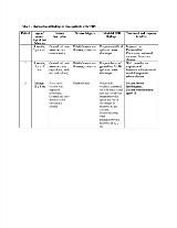
Rationale: Wolf-Hirschhorn Syndrome (WHS), an extremely rare genetic syndrome caused by deletion of a portion of the short arm of chromosome 4p, is characterized by many congenital abnormalities including distinctive “Greek helmet” facies, microcephaly, and cardiac defects with developmental delay, intellectual disability, and seizures. Epilepsy reportedly affects 50-100% of WHS patients and likely results from the deletion of a seizure susceptibility region on chromosome 4p. Currently, however, little information is available regarding the associated seizure phenotypes, electroencephalographic (EEG) features and seizure/epilepsy outcomes. This case series highlights the seizure phenotypes and EEG findings in three patients with WHS. Methods: A retrospective chart review of three patients with a known diagnosis of WHS was conducted and data was extracted using electronic medical records. We aimed to assess the age of seizure onset, seizure types, EEG findings and response to treatment with anti-epileptic drugs (AEDs). Results: Clinical Features:Each patient exhibited phenotypic abnormalities consistent with WHS - “Greek helmet” facies (orbital hypertelorism, a prominent glabella, and a flattened nasal bridge), seizures, developmental delay, and cardiac defects. Additional features common to the syndrome included failure to thrive and hypotonia (two patients), urinary tract malformations (one patient) and a rare immunodeficiency with cyclic fevers (one patient). Electroclinical Findings:Analysis of the seizure phenotypes, EEG results and response to AEDs are summarized in Table 1. Conclusions: This small observational study of three patients highlights the electroclinical patterns of epilepsy seen in WHS. Variable seizure semiology, onset within the first year of life with special susceptibility to febrile and intercurrent illnesses causing seizures to cluster were common findings in all our patients. Additionally, as seen in one of our patients, associated immunodeficiencies in WHS predisposes to an increased number of infections, especially within the first few years of life, which may further compound the seizure burden. Consequently, similar to patients with simple febrile seizures, educating these families and providing specific instructions on anti-pyretics and anti-epileptic prophylaxis during sick periods is useful in ameliorating the seizure burden. Just as there is no one seizure phenotype, no single EEG pattern is pathognomonic for WHS. However, especially of interest and somewhat unique seem the fast spike–polyspike-wave complexes over the posterior brain regions triggered by eye closure. This was seen in one of our patients and has been reported in literature. Overall, despite the limitation of our small sample size, seizures seemed to be well controlled with typical AEDs such as levetiracetam, phenobarbital, and valproic acid depending on the age and seizure type. Funding: None
