AFTER DISCHARGE (AD) STIMULATION ON SIMULTANEOUS MEG AND INTRACRANIAL EEG (ECoG) AND SPIKE SOURCE RECONSTRUCTION
Abstract number :
3.151
Submission category :
Year :
2005
Submission ID :
5957
Source :
www.aesnet.org
Presentation date :
12/3/2005 12:00:00 AM
Published date :
Dec 2, 2005, 06:00 AM
Authors :
1Dinah Thyerlei, 1Nancy Lopez, 2Adam N. Mamelak, and 1,2William W. Sutherling
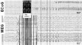
Cortical stimulation of intracranial grid electrodes (ECoG) is used to identify functionally relevant areas before brain surgery. We recorded ECoG stimulation and simultaneous magnetoencephalogram (MEG). Stimulating an intracranial electrode causes a local high current directly under the contact, which may cause AD and thereby provides a known current source location at the patient[apos]s cortex. We reconstructed the spike source from both ECoG and MEG and compared the results to the true location of the stimulated grid contacts. Stimulation to identify functionally important areas and to determine AD threshold was performed with symmetric biphasic pulses for 0.3 ms, with a 50 Hz frequency and 2s trains. The current intensity was increased every 20 s starting at 1 mA up to 15 mA or until AD occurred. 64 channel ECoG and 68 sensor whole-head MEG (VSM-CTF) were recorded simultaneously. The electrode positions were taken from CT and MRI. Multiple unaveraged early AD spikes seen simultaneously in only the two stimulated intracranial electrodes and on MEG sensors were used to calculate the spike sources by using different head and source models. Localization accuracy was compared between found sources of the different models and the known localization of the two stimulated electrodes. If only the earliest fronto-temporo-parietal AD spikes with no propagation were used and the spikes were visible on the MEG sensors without averaging, the mean distance between MEG spike sources and the center of the two stimulated electrodes was 7.8 mm (SD 1.7) for a single sphere and ECD. Using RAP-MUSIC with a fit window of 50 ms around the spike peaks yielded similar distances. Surface-constrained minimum norm solutions found more widespread activity. For ECoG the mean distance between the ECoG source and center of the stimulated electrodes was 3.7 mm (SD 1.4). Source localization of AD spikes seen on subtemporal strips and MEG yielded larger distances. Co-registration of ECoG AD stimulation and whole-head MEG allows a direct evaluation of different reconstruction methods. Only non-propagated AD spikes on ECoG should be chosen for source analysis. The accuracy of MEG source localization for AD spikes for the simplest model (ECD and single sphere) is good. Postsurgical alterations (ECoG/cable artifact, edema) influence the correct localization of ECoG electrodes on CT/MRI.[figure1] (Supported by NIH grants NINDS RO1-NS20806, NCRR S10RR13276, NIHM MH53213, and the Norris, Zeilstra and Bacon Foundations.)
