ALTERATIONS IN GLIAL GLUTAMATE UPTAKE AND METABOLISM IN AREAS OF SCLEROSIS IN TEMPORAL LOBE EPILEPSY
Abstract number :
3.041
Submission category :
Year :
2002
Submission ID :
3409
Source :
www.aesnet.org
Presentation date :
12/7/2002 12:00:00 AM
Published date :
Dec 1, 2002, 06:00 AM
Authors :
Alexander A. Sosunov, Robert R. Goodman, Guy M. McKhann II. Neurological Surgery, Columbia University, New York, NY
RATIONALE: Astrocytes are critically important regulators of glutamate in the central nervous system, clearing neuronally released glutamate from the extracellular space and subsequently cycling glutamate to glutamine via the enzyme glutamine synthetase or converting glutamate for utilization in the TCA cycle via the enzyme glutamate dehydrogenase. To test the hypothesis that glutamate uptake and metabolism are impaired in areas of hippocampal sclerosis in human temporal lobe epilepsy, we carried out immunohistochemical analysis of glutamine synthetase, glutamate dehydrogenase, and astrocytic glutamate transporter proteins in resected human hippocampi.
METHODS: Fifteen hippocampi obtained from temporal lobe epilepsy surgeries were studied. Double labeling immunohistochemistry combined with confocal microscopy was used to evaluate the neuroglial capacity for uptake and metabolism of glutamate.
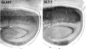
RESULTS: As detected by MAP-2 staining, all cases had hippocampal sclerosis, with marked neuronal loss in the CA1, CA3, and hilar regions. Astrocytes in these sclerotic areas had high levels of GFAP and S100b expression, in contrast to very weak immunoreactivity for glutamine synthetase (GS). In contrast, in less sclerotic regions (subiculum, CA2, molecular layer of dentate gyrus) high levels of astrocytic GS immunoreactivity were detected, with lower expression of GFAP and S100b. Figure 1 demonstrates representative immunostaining of human epileptic hippocampus showing increased GFAP and decreased GS expression in areas of sclerosis. No difference was seen in GDH expression in sclerotic and nonsclerotic areas of hippocampus. Immunoreactivity for the astrocytic glutamate transporters GLAST and GLT-1 was visualized in nonsclerotic regions but markedly decreased in areas of sclerosis (Figure 2). Unlike astrocytes, oligodendrocytes displayed high levels of GS immunoreactivity in sclerotic areas but were GS negative in nonsclerotic regions. S100b expression was observed in oligodendrocytes only in nonsclerotic areas.
CONCLUSIONS: In areas of hippocampal sclerosis, astrocytes express low levels of glutamate transporters and glutamine synthetase, suggesting that glutamate clearance and metabolism is impaired in these regions. There may be compensatory glutamine synthesis by oligodendrocytes in sclerotic tissue.
Objective: Participants should be able to describe glial alterations in glutamate uptake and glutamine synthesis in areas of sclerotic hippocampus.[figure1][figure2]
[Supported by: the Klingenstein Foundation, New York Academy of Medicine, and Parents Against Childhood Epilepsy (P.A.C.E) (GM).]
