An Interesting Case of a Low-Grade Glioma Masquerading as Generalized Epilepsy
Abstract number :
3.330
Submission category :
9. Surgery / 9B. Pediatrics
Year :
2018
Submission ID :
501219
Source :
www.aesnet.org
Presentation date :
12/3/2018 1:55:12 PM
Published date :
Nov 5, 2018, 18:00 PM
Authors :
Nitin Agarwal, Minnesota Epilepsy Group, P.A.; Wenbo Zhang, Minnesota Epilepsy Group, P.A.; and Meysam Kebriaei MD, Children's Minnesota
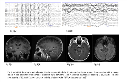
Rationale: Seizures are a common presentation of pediatric brain tumors with neuroglial tumors and gliomas being the most commonly associated ones. Tumor related seizures are mostly focal, with or without secondary generalization, and true generalized seizures have been rarely reported. Other focal lesions are known to present with generalized EEG findings. Methods: We report an interesting case of a 6-year-old male presenting with generalized seizures and epileptic spasms in the setting of brain tumor. Results: A 6-year-old right-handed male, born and brought up in Vietnam, was referred to our center for a second opinion due to refractory epilepsy. He had 2 unclear episodes of altered consciousness at 9 months and 2 years of age respectively and was subsequently diagnosed with epilepsy at 5 years of age before moving to United States. Seizures were described as staring off with a scared look on the face, crying facies, bilateral arms raised upwards with clawed hands and tense elbows. This occurred in clusters over a 5-10 min. period and occurred on average 1-2 times per day at the presentation. Previous EEGs noted multifocal and generalized epileptiform discharges and surgical treatment was ruled out. He was being treated with 4 anti-epileptic medications (AEDs) and had failed 4 others in the past.Pre-surgical EEG evaluation showed multifocal (maximum right and left temporal, at times independent) epileptiform discharges along with generalized polyspikes (Fig 1a). Typical seizures were captured and were consistent with myoclonic jerks, axial tonic seizures and epileptic spasms (Fig 1b). Brain MRI showed cystic lesion in the inferior-medial aspect of the right temporal lobe involving para-hippocampal gyrus, measuring 2.0 x 1.4 x 1.1 cm with ill defined T2/FLAIR hyper intensity surrounding the cyst (Fig 1c-f). Magnetoencephalography showed dipoles in right frontal, right temporal and left temporal lobe. Neuropsychological evaluation showed mild to moderate cognitive delays encompassing both verbal and non-verbal abilities with mild-moderate intellectual disability.After review in the patient management conference, it was decided to proceed with the surgery without further invasive evaluation. Surgery included resection of the right temporal lesion and mesial temporal lobe. Biopsy of the lesion confirmed WHO grade I, low-grade neuroepithelial neoplasm. Post-resection EEG at 2-months interval showed complete resolution of epileptiform activity and only focal slowing was noted in the right temporal region (Fig 2). At the last clinical follow up at 8-month interval, he remains completely seizure free with 50% reduction in his medications. Interval neuropsychology testing showed stable verbal and non-verbal cognitive development with improvement in clarity and quantity of speech and in social relatedness. Conclusions: This case highlights the fact that brain tumors may present with predominant generalized seizures and EEG findings but does not preclude surgery. Complete seizure freedom can be achieved even without invasive EEG evaluation in carefully selected cases. Funding: None

.tmb-.png?Culture=en&sfvrsn=6eb6d66_0)