AUTOMATIC SPIKE DETECTION FOR SOURCE LOCALIZATION IN CHILDREN AND NEONATES
Abstract number :
2.171
Submission category :
Year :
2004
Submission ID :
4693
Source :
www.aesnet.org
Presentation date :
12/2/2004 12:00:00 AM
Published date :
Dec 1, 2004, 06:00 AM
Authors :
1Ardalan Aarabi, 1Nadege Roche Labarbe, 1Arnaud Montigny, 2Guy Kongolo, 2Patrick Berquin, 3Michiel Van Burik, 1Reinhardt Grebe, and 1,2Fabrice Wallois
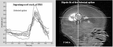
Interictal spikes, in children, and Positive Rolandic Spikes (PRS), in preterms, are transient abnormal activities indicating specific pathologies (i.e. epilepsy and Periventricular Leukomalacia (PVL)). EEG source localization gives new insights for identification of the brain area where focal activities originate. Spike shape classification is essential for good dipole fit accuracy.We designed an automated method that extracts spikes and detects the clusters of neuronal activities for EEG source localization in epileptic children and preterms. We used template matching to extract spikes to discriminate individual neuronal activities. The desired spike is captured as the reference spike template, then all spike candidates are scanned for waveforms matching the template within a specified error range. The sum of squared error (SSE) is used as a criterion for the goodness of fit. When SSE reaches a value below the preset detection-threshold, detection is reported. To track changes in the shape of the spikes, matched spikes are averaged during template matching in order to adapt the reference spike template. The above mentioned method is applied to spontaneous EEG data sets, collected from epileptic children (64 channels) and preterm (21 channels) with PVL. The Dipole Fit method (ASA, ANT Software) is used to localize the equivalent dipoles for all extracted spikes. The spatial topography associated with the sources was calculated using a realistic head model. Spike clustering was carried out in temporal and spatial domains for interictal and PRS activities. The source localization shown below resulted from our spike clustering method in a newborn with PRS. In this case, mean and variance of SSE were 0.5886 and 0.1460 for the selected spikes, and respectively 0.7167 and 0.1854 for the rejected ones. Then, the localized dipoles were selected to be in the same cluster by comparing the distance between the center of cluster and dipoles with the predetermined threshold.[figure1] Lack of a unique definition for specific age-dependant cerebral activities (normal: delta brush, frontal transients, and abnormal: PRS) in preterm makes the template matching method very useful in extracting the spikes. This method allows a better discrimination between different sources of activities by using contextual information. (Supported by French Ministry of Research, Region Picardie, University of Picardie (HTSC Project [amp] ACI [quot]Integrative and Computational Neurosciences[quot] Project).)
