Background EEG Activity Differentiates Between Physiological and Epileptic High-Frequency Oscillations and Remains Unaltered Over Several Days
Abstract number :
2.007
Submission category :
3. Neurophysiology / 3A. Video EEG Epilepsy-Monitoring
Year :
2018
Submission ID :
501083
Source :
www.aesnet.org
Presentation date :
12/2/2018 4:04:48 PM
Published date :
Nov 5, 2018, 18:00 PM
Authors :
Annika Minthe, University of Freiburg; Daniel Lachner Piza, University of Freiburg; Wibke Janzarik, University of Freiburg; Peter Reinacher, University of Freiburg; Matthias Dümpelmann, University of Freiburg; and Julia Jacobs, University of Freiburg
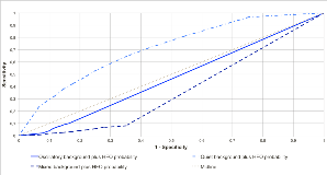
Rationale: High-frequency oscillations (HFO) are markers of epileptic tissue. Recently different HFO patterns were described depending on the EEG background activity. HFO patterns with continuously oscillating background were found to be physiological, while HFO arising from quiet background were linked to epileptic tissue. HFO-Background pattern analyses therefore allowed distinguishing physiological and epileptic HFO activity. It remains unclear whether the EEG background activity in the high-frequency band is subject to alterations and the stability of HFO pattern over several days and different sleep-wake stages has not been investigated yet. We hypothesize that epileptic HFO patterns remain stable over several days in time during the chronic intracranial EEG investigation. Methods: HFO patterns (oscillatory vs. quiet background, Figure 1) were analysed in 23 consecutive patients implanted with depth and subdural grid electrodes in the Epilepsy Center Freiburg. Pattern scoring was performed on every channel in 10 second intervals of 2 minutes in three day- and night-time EEG-segments for every patient. An entropy value, which measures variability of patterns per channel, was calculated. High entropy values would suggest frequent changes in background activity. Differences in pattern probability and entropy were calculated for inside and outside seizure onset (SOZ), different electrode types and brain regions. Results: 2089 channels with 11940 EEG segments were analysed. We found strong association between areas with sporadic short HFO out of a flat baseline activity and SOZ-channels (Figure 2). HFOs deriving from continuous oscillatory activity seemed to be randomly represented in some channels. Entropy values between EEG segments were significantly lower in channels with flat baseline plus HFO and within the SOZ compared to areas with oscillatory background (0.57 ± 0,39 vs. 0,72 ± 0,37; p < 0,001). Conclusions: As has been suggested before HFO in a quiet background were strongly linked to the SOZ while HFO from an oscillatory background were not and might be physiological. HFO background patterns remained stable over several days and during night and daytime recordings. Our findings suggest that interaction between HFO and background activity are a stable phenomenon reflecting different mechanisms of underlying brain activity. HFO pattern analysis therefore might be a reliable way of identifying epileptic and physiologically active brain regions after looking at very short time segments. Funding: German Research Foundation (grant no.:JA 1725/2-1)

.tmb-.png?Culture=en&sfvrsn=6fb2f55c_0)