CAN dSPM BASED ON MEG BE A NEW METHOD TO SHOW PRIMARY AREA AND PROPAGATION FOR BILATERAL PARTIAL EPILEPSY?
Abstract number :
2.323
Submission category :
Year :
2004
Submission ID :
4772
Source :
www.aesnet.org
Presentation date :
12/2/2004 12:00:00 AM
Published date :
Dec 1, 2004, 06:00 AM
Authors :
K. Hara, F. Lin, S. Camposano, D. Foxe, P. E. Grant, and S. M. Stufflebean
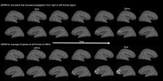
Patients (pats) with partial epilepsies (PE) may candidates for surgery. In pats whose EEGs show bilateral interictal spikes (IIS), it is necessary to precisely localize their primary epileptogenic area before surgery. We report a use of dynamic statistical parametric modeling (dSPM) and Magnetoencephalography (MEG) to investigate primary area and propagation in PE showing bilateral IIS on electroencephalograly (EEG). Pats with refractory PE with bilateral EEG IIS were studied using 306-channel MEG (Neuromag) and 70-channel EEG for [sim]60mins including sleep. Equivalent current dipoles (ECD) were calculated for MEG IIS. Only ECDs with Goodness of Fit [gt]80% and 100[lt]Qvalue[lt]400nAm were accepted. The MEG dSPM were calculated. There are 7 pats who showed bilateral independent EEG IIS. 3 of 7 showed 3-8 IIS propagated from one to another region over short durations on MEG (15ms[sim]382ms). There were no propagations in the opposite direction. We suspected the first region prior to propagation showed the primary area. To confirm this, we calculated average dSPM for a pat.
Sample; An11 year old girl. EEG showed independent left and right IIS (more spikes at left F3 than right) on EEG using 2 kinds of bipolar montage and a monopolar montage. Some MEG IIS were not accompanied by EEG IIS, and some right MEG IIS appeared at left on EEG. MEG showed 3 types IIS, 8 IIS clustered at right frontal (Clu R), 16 IIS at left frontal (Clu L) and 3 IIS propagated from right frontal to left frontal region (Clu RL). We suspected that all left frontal IIS were propagated from the right frontal area. To support our assumption, we calculated the dSPM of the averaged spikes for each spike cluster. The dSPM of the average spikes in Clu RL and even dSPM for each 3 IIS in Clu RL showed clear propagation from right to left frontal lobe. (Fig. Upper pictures) In Clu L, the dSPM of each spike did not showed clear propagation. However the dSPM of the average spikes in Clu L showed the right frontal activity proceeding to left frontal lobe activity. (Fig. Lower pictures) The duration between right and left frontal activity (24ms) and locations in the left and right frontal lobe were same as each and average dSPM of Clu RL. The dSPM of average for Clu R did not show proceeding activity. This result supports our assumption that the left frontal activity is propagated from a right frontal focus.[figure1] In cases with bilateral independent IIS on EEG, the MEG and dSPM is useful delineate the primary region and the existence of propagation from one to anther region. MEG with dSPM may be useful for presurgical evaluation. (Supported by The MIND Institute)
