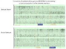
Cessation of gamma activity in the centromedian nuclei associated with spread of focal seizures to the anterior nuclei of the thalamus
Abstract number :
3.289
Submission category :
Late Breakers
Year :
2013
Submission ID :
1843312
Source :
www.aesnet.org
Presentation date :
12/7/2013 12:00:00 AM
Published date :
Dec 5, 2013, 06:00 AM
Authors :
B. A. Leeman-Markowsk, O. Smart, R. Faught, R. Gross, K. Meador
Rationale: Impairment of consciousness during seizures may be mediated by propagation of ictal activity to the thalamus. The precise thalamic regions involved and their roles with respect to maintenance of consciousness, however, are unclear. The centromedian (CM) nucleus of the thalamus likely plays a role in arousal, given its connections within the ascending reticular activating system. We propose that alterations of firing patterns within the CM during ictal activity, seen in the following case presentation, contribute to impaired arousal during focal seizures.Methods: EEG data were collected from a patient who underwent intracranial monitoring for clinical purposes, which included placement of bilateral depth electrodes within the CM and anterior nuclei (AN) of the thalamus. Spectral power was computed across ictal state (preictal, ictal, and postictal) and level of consciousness (stupor/sleep vs. awake) in the CM and AN using spectrograms (1-sec window, 75% overlap, Goertzel Discrete Fourier Transform algorithm) of differential electrode signals in each thalamic nucleus. We compared gamma (30-40 Hz) power for combinations of ictal state, level of consciousness, and nucleus using a Kruskal-Wallis ANOVA and Mann Whitney Wilcoxon tests.Results: Over 100 seizures were captured, characterized by an atonic slump of the head, left arm flexion, and loss of consciousness. Seizures had multifocal onsets from the left temporal and frontal regions, although multiple seizures were non-localizable. Ictal activity typically spread to the left AN, often involving the right AN as well. At baseline, the bilateral CM demonstrated rhythmic bursts of gamma activity, evident more often and with greater amplitude during wakefulness. This activity ceased as ictal discharges spread to the AN and consciousness was impaired, and it recurred with the termination of each seizure as the patient regained awareness (Figure 1). Gamma power was analyzed for a subset of four seizures, demonstrating statistical significance of this CM pattern (Figure 2). When seizures occurred during wakefulness, there was lower CM ictal power and higher CM postictal power relative to the preictal epoch. As the seizures spread to the AN, there was higher ictal AN power with lower AN postictal power relative to the preictal epoch (p<0.0001, all pairings). In contrast, this spectral pattern was not evident when seizures occurred during sleep, with higher ictal and lower postictal power in both the CM and AN relative to the preictal epoch (p<0.0001, all pairings).Conclusions: The data revealed a characteristic pattern of gamma bursts in the CM during wakefulness. Cessation of the bursts during partial seizures was associated with impaired consciousness, consistent with connectivity, lesion, neuroimaging, and electrical stimulation studies which suggest that the CM participates in regulation of vigilance and arousal. Alternative explanations could include CM s role in motor activity.
