Characteristics of Interictal Slow Wave Activity From Bilateral Invasive EEG During Sleep and Postictal States
Abstract number :
2.024
Submission category :
3. Neurophysiology / 3B. ICU EEG
Year :
2018
Submission ID :
502411
Source :
www.aesnet.org
Presentation date :
12/2/2018 4:04:48 PM
Published date :
Nov 5, 2018, 18:00 PM
Authors :
Brian N. Lundstrom, Mayo Clinic; Melanie Boly, University of Wisconsin; Robert Duckrow, Yale University School of Medicine; Hitten P. Zaveri, Yale University; and Hal Blumenfeld, Yale University School of Medicine
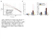
Rationale: Increased slow wave activity is typically associated with decreased levels of awareness. Previous work has shown greater slow wave activity (1-2 Hz) in frontal and parietal areas during seizures that impair awareness than during seizures that do not [1]. During non-REM sleep, slowing was increased for patients with epilepsy compared to patients without epilepsy, including a local increase at the seizure onset zone (SOZ) [2]. Here, we look at the spatial distribution of slow wave activity relative to the SOZ in epilepsy patients, and compare sleep and postictal states. Methods: Data were recorded with the Natus Neurolink IP 256 channel EEG amplifier (0.16 Hz High pass filter, 1024 Hz sampling frequency). Inclusion criteria were: (1) bilateral subdural electrode coverage (>30 electrode contacts per hemisphere), (2) no known hemispheric abnormalities or diffuse abnormalities affecting the frontal lobes and (3) well-defined SOZs. Intracranial data from eight patients with 196, 172, 203, 201, 199, 213, 154, and 223 subdural contacts, respectively. 1061 contacts were ipsilateral to the SOZ and 500 contralateral. SOZs were located in the left frontal (n =4), left temporal (n = 1), right temporal (n = 3), and right parietal/occipital (n = 1) lobes. One patient had two SOZs (right temporal and right parietal/occipital). 15 minutes in the wake, slow wave sleep, and postictal states following each of three focal seizures was examined for five patients, with three epochs of wake and slow wave sleep each for the remaining three patients. Results: Postictal slowing is characterized by a broadband power increase near the SOZ, whereas sleep shows an increase in delta (0.3-4 Hz) but not beta/gamma power compared to wakefulness (figure 1). Relative slow oscillation (0.3-1 Hz) power is decreased close to the SOZ and increases with distance from the SOZ (figure 2). Contacts near the SOZ show decreased spatial correlations as well as decreased coupling between slow oscillations and beta/gamma frequencies during sleep. Conclusions: Power less than 1 Hz is decreased near the SOZ and may represent decreased efficacy of inhibitory mechanisms. Beta and low gamma frequency power is increased close to the SOZ, perhaps related to interictal epileptiform activity. Decreased slow delta (0.3-1 Hz) to beta and low gamma power, as evidenced by a counter-clockwise tilt of the power spectrum, may represent a signature of hyper-excitable tissue. The broadband power increase during the postictal state suggests an overall increase of neural unit activity contributing to the EEG signal. In contrast, sleep shows an increase primarily in slower frequencies, as can be seen with a change in firing patterns. Further results suggest that the hemisphere ipsilateral to the SOZ has a reduced capacity for spatial and temporal synchrony. Overall, there are distinct interictal differences near the SOZ, which may be more apparent during sleep than postictal states. References: Englot et al. Impaired consciousness in temporal lobe seizures: role of cortical slow activity. Brain. 2010;133(Pt 12):3764-377.Boly et al. Altered sleep homeostasis correlates with cognitive impairment in patients with focal epilepsy. Brain. 2017;140(4):1026-1040. Funding: Mayo Clinic Scholar Program

.tmb-.png?Culture=en&sfvrsn=9202cb7f_0)