COINCIDENTAL MRI CHANGES IN TEMPORAL PARIETAL OCCIPITAL CORTEX AND THE IPSILATERAL PULVINAR ASSOCIATED WITH PARTIAL STATUS EPILEPTICUS
Abstract number :
2.324
Submission category :
Year :
2004
Submission ID :
4773
Source :
www.aesnet.org
Presentation date :
12/2/2004 12:00:00 AM
Published date :
Dec 1, 2004, 06:00 AM
Authors :
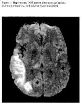
J. Craig Henry, and Michel J. Berg
Reversible diffusion-weighted MR imaging (DWI) hyperintensities have been reported in the setting of status epilepticus, and likely represent seizure-induced cytotoxic edema. We describe a neuroanatomically distinct pattern of cortical and thalamic DWI changes among six patients with partial status epilepticus. The history, EEG findings and serial neuroimaging results of six patients with partial status epilepticus and focal, reversible DWI hyperintensities on MRI were reviewed and compared. While DWI abnormalities were present in all six patients in the series, two of three patients with predominantly posterior temporal, parietal and occipital involvement had concomitant reversible DWI hyperintensities in the ipsilateral pulvinar. No thalamic DWI abnormalities were found in patients whose MRI changes involved primarily frontal cortex.[figure1] The pattern of MRI findings in this series suggests involvement of the pulvinar with seizures that emanate primarily from posterior temporal, parietal or occipital cortices. A similar coincidence of cortical and thalamic MRI abnormalities is reflected in previous reports, though not specifically identified. Anatomically, the pulvinar receives afferents from the superior and inferior parietal lobules, posterior temporal region, and peristriate occipital cortex. Our neuroimaging findings reinforce the functional significance of these connections, and comprise a distinct MRI pattern in temporal-parietal-occipital partial status epilepticus.
