Comparison of P3 responses before and after surgery in TLE patients using CD analysis
Abstract number :
2.382
Submission category :
18. Late Breakers
Year :
2010
Submission ID :
13439
Source :
www.aesnet.org
Presentation date :
12/3/2010 12:00:00 AM
Published date :
Dec 2, 2010, 06:00 AM
Authors :
A. Matsuda, K. Hara, M. Miayajima, K. Ohta, T. Maehara, E. Matsushima, M. Watanabe, M. Hara, M. Matsuura
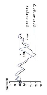
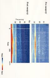
Rationale: To better characterize changes in cognitive function resulting from surgery, EEG was recorded in patients with temporal lobe epilepsy (TLE) before and after surgery as they performed an oddball task. Two types of analyses compared function pre- and post-surgery: event-related potential (ERP) analyses focusing on the P3 component, and a frequency analysis using complex demodulation (CD).Methods: Five TLE patients (2 females, mean age 30.1 6.5 years, all right handed) participated in the study. Patients took a various number of anti-epileptic drugs (AED), with two patients taking one AED, two patients taking two AEDs, and one patient taking three AEDs. Seizure frequency prior to surgery also varied such that it was daily in one patient, weekly in two patients and monthly in two patients. All patients were seizure free after surgery. Epileptic foci were localized to the left hemisphere for two patients and to the right hemisphere for three patients. All patients were hospitalized for pre-surgical monitoring, epilepsy surgery and post-operative controls in the Tokyo Medical and Dental University Medical Center. Pre-surgery examinations were conducted one to two months prior to surgery (mean = 28.0 days), and post-surgery examinations were conducted between one to four months after surgery (mean = 42.8 days). EEG was recorded during an auditory oddball task from four midline locations (Fz, Cz, Pz and Oz). Stimuli consisted of 1000Hz pure tones (frequent; 80% of trials) and 1050Hz tones (rare; 20% of trials). A minimum of 20 good deviant trials were included in each participant s average waveform. Participants were instructed to count the number of rare stimuli while watching silent cartoons. For ERP analyses, peak amplitude, mean amplitude and peak latency were evaluated at each electrode 400-530ms post-stimulus onset. Frequency analyses were performed using CD. Induced EEG responses were first divided into 0.25 Hz bins (ranging from 0 50 Hz), and amplitude was then evaluated for each bin. EEG and EOG (electrooculographic) data were filtered from 0 50 Hz. Statistical analyses included two-way repeated measures ANOVAs. Results: P3 grand average waveforms are shown in Fig. 1. There was no significant difference among them. CD analyses of induced EEG responses revealed that delta ( -3 Hz) and theta (4-7 Hz) activity increased after surgery, whereas fast activity (25-40 Hz) decreased (shown in Fig. 2).Conclusions: Although there were no significant differences in the pre- and post-surgery P3 grand averages, CD analyses revealed increased delta and theta activity as well as decreased fast activity after surgery. CD analysis may thus be a useful method for observing cognitive function.

