Cortical Thinning in Epilepsy Patients With Postictal Generalized EEG Suppression
Abstract number :
2.171
Submission category :
5. Neuro Imaging / 5A. Structural Imaging
Year :
2018
Submission ID :
501416
Source :
www.aesnet.org
Presentation date :
12/2/2018 4:04:48 PM
Published date :
Nov 5, 2018, 18:00 PM
Authors :
Yingying Tang, West China Hospital of Sichuan University; Dongmei An, West China Hospital of Sichuan University; Yuan Xiao, West China Hospital of Sichuan University; Running Niu, West China Hospital of Sichuan University; Xin Tong, West China Hospital of
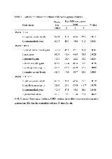
Rationale: To investigate the brain microstructural abnormalities in epilepsy patients with postictal generalized electroencephalographic suppression (PGES) using a cortical surface-based analysis. Methods: According to the video-electroencephalography records of epilepsy patients with generalized convulsive seizures (GCSs), we recruited 30 patients with PGES (PGES+) and 21 patients without PGES (PGES-). High-resolution T1-weighted images were acquired from each patient and 30 matched healthy control subjects (HCs). Cortical thickness was compared among the three groups using FreeSurfer software. Results: Patients with PGES showed reduced cortical thickness in the right paracentral lobule, inferior parietal lobule, supramarginal gyrus (SMG) and middle temporal lobe compared with patients without PGES. In relation to HCs, the PGES+ group presented reduced cortical thickness in the right superior parietal lobule (SPL) and SMG, while the PGES- group presented reduced cortical thickness in the left precuneus, precentral gyrus, lateral occipital gyrus, parahippocampal gyrus, SPL, and right caudal middle frontal gyrus. Conclusions: Patients with PGES exhibited characteristic brain microstructural abnormalities, corroborating the PGES mechanisms at the brain level. The right-side predominance of the detected PGES-related cortical thinning was the same as that of SUDEP cases and patients at high risk for SUDEP, implying that PGES and SUDEP may share a common abnormal brain substrate that is involved in the pathophysiology of these conditions. Funding: This study was supported by the National Natural Science Foundation of China (Grant No. 81601133, 81420108014, and 81771402).
