DECREASED GLUTAMINE SYNTHESIS IN HUMAN TEMPORAL LOBE EPILEPSY: CHICKEN OR EGG?
Abstract number :
1.061
Submission category :
Year :
2004
Submission ID :
4162
Source :
www.aesnet.org
Presentation date :
12/2/2004 12:00:00 AM
Published date :
Dec 1, 2004, 06:00 AM
Authors :
1Guy M. McKhann II, 1Xiaoping Wu, 1Robert R. Goodman, 2Peter D. Crino, and 1Alexander A. Sosunov
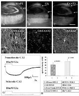
Deficiency in glutamine synthetase (GS), the astrocytic enzyme that converts glutamate to glutamine, was reported as an explanation for elevated extracellular glutamate levels and seizure initiation in mesial temporal lobe epilepsy (MTLE). We studied astrocytes from resected human TLE to determine whether alterations in GS are a primary etiological factor or are secondary to existing alterations in the epileptic hippocampus. Resected human hippocampi from MTLE (n=11) and non-MTLE (n=6) patients were studied by a combination of quantitative immunohistochemistry and acute hippocampal slice electrophysiology. We investigated glutamate uptake and glutamine synthesis by astrocytes in the CA1 subfield in these two pathologies. Astrocytes from non-MTLE epileptic hippocampi express a high level of glutamate transporters and GS (Figure 1D,H), and demonstrate inward transporter currents in response to glutamate application (Figure 1G). In contrast, in areas with prominent neuronal loss and astrogliosis in the MTLE epileptic hippocampus, there is markedly decreased expression of both of the astrocytic glutamate transporters, EAAT1 (not shown) and EAAT2 (Figure 1E,H). This decrease parallels the observed downregulation of GS in these areas (Figure 1B,E,H). There is little to no inward glutamate induced current in individual astrocytes recorded in areas of sclerosis in acute hippocampal slices from MTLE patients (Figure 1G).[figure1] Our results demonstrate that there is decreased glutamate transporter expression together with impaired glutamate uptake by astrocytes in areas of sclerosis in MTLE. These findings suggest that downregulation of GS in MTLE is a secondary phenomenon in response to glutamate not entering these cells, rather than a primary enzymatic defect. Defective glial glutamate uptake has also been implicated in astrocytic tumor growth, neurotoxicity, and seizures.
[underline]Legend to Figure 1.[/underline]
A-C.Confocal images are shown of: A) Immunohistochemical staining with the neuronal marker MAP2 from a patient with MTLE. There is little expression of GS (B) or EAAT2 (C) in astrocytes in areas of neuronal loss (arrows in B,C delineate the CA1 region). D-E. Immunolabelling for GS (red) and EAAT2 (green) from non-MTLE (D) and MTLE (E) epileptic hippocampi (CA1). Scale bars: A,B,C [ndash] 1.5 mm; D,E,F [ndash] 80 mm. G. Astrocytes recorded from non-MTLE and MTLE CA1 subfields. H. Quantitative immunohistochemistry. (Supported by Klingenstein Foundation (GM)
NIH R21 NS 42334 (GM, PC)
New York Academy of Medicine Elsberg Fellowship (GM))
