DENTATE GRANULE CELL DISINHIBITION AND HYPEREXCITABILITY IN AWAKE RATS DURING THE FIRST WEEK AFTER KAINATE-INDUCED STATUS EPILEPTICUS; THE SOURCE OF VARIABILITY IDENTIFIED
Abstract number :
E.02
Submission category :
Year :
2002
Submission ID :
1463
Source :
www.aesnet.org
Presentation date :
12/7/2002 12:00:00 AM
Published date :
Dec 1, 2002, 06:00 AM
Authors :
Brian D. Harvey, Robert S. Sloviter. Departments of Pharmacology and Neurology, University of Arizona College of Medicine, Tucson, AZ
RATIONALE: Status epilepticus (SE) in rats produces widespread brain damage, variable patterns of hippocampal cell loss, mossy fiber sprouting (MFS), and spontaneous seizures of unknown origin. We reported that granule cell disinhibition and hyperexcitability developed immediately after kainate (KA)-induced SE, and were closely correlated with hilar cell loss. Restoration of inhibition, rather than hyperexcitability, coincided with MFS (1). Our suggestion that MFS is inhibitory rather than epileptogenic was addressed in a recent study which concluded that inhibition recovers in the first days after SE, prior to MFS (2).
METHODS: Rats were implanted bilaterally with stimulating electrodes in the perforant pathway and recording electrodes in the granule cell layers. SE lasting 6-8hr was reliably induced in awake rats by KA (10mg/kg iv; n=13). Spontaneous and evoked granule cell activity were recorded and histological analysis was subsequently performed.
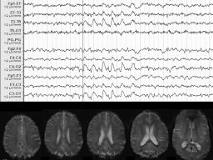
RESULTS: Rats varied in terms of the time spent generating granule cell discharges during KA-SE. Rats exhibiting granule cell discharges ~65% of the time during behavioral SE (n=5) immediately exhibited multiple granule cell population spikes and paired-pulse disinhibition. Subsequent histological analysis showed extensive hilar cell loss and dense MFS. Conversely, rats exhibiting similar behavioral SE, but discharging only 10-30% of the time (n=8) exhibited single or double population spikes, minimal granule cell disinhibition, minor hilar cell loss, and minimal MFS. The latter group exhibited rapid restoration of paired-pulse inhibition, whereas the former exhibited a prolonged disinhibitory state throughout the week after SE. Spontaneous population spikes, multiple evoked spikes, and long-latency discharges often attributed to MFS in bicuculline-treated hippocampal slice preparations were observed in awake rats before MFS (figure below).
CONCLUSIONS: We attribute the rapid recovery of paired-pulse inhibition in minimally affected animals to a compensatory mechanism operant in relatively normal tissue. In rats with extensive hilar cell loss, granule cell pathophysiology did not recover before MFS. Thus, we attribute the rapid recovery of inhibition in the study of Hellier et al (2) to the fact that none of their 17 animals exhibited the degree of multiple spiking and disinhibition described in our previous (1) or present studies. We conclude that despite similar behavioral SE among animals, granule cell discharges and hilar cell death occur unreliably and unpredictably. When granule cell discharges occur consistently during KA-SE, hilar cells die, prolonged disinhibition results immediately, and the restoration of inhibition corresponds temporally with MFS.
1. Sloviter RS (1992) Neurosci Lett 137:91-96.
2. Hellier JL, Patrylo PR, Dou P, Nett M, Rose GM, Dudek FE (1999) J Neurosci 19:10053-10064.[figure1]
[Supported by: This work was supported by NINDS grant NS18201.]
