DEVELOPMENT OF ATROPHY IN SUPER REFRACTORY STATUS EPILEPTICUS
Abstract number :
2.371
Submission category :
Year :
2014
Submission ID :
1868923
Source :
www.aesnet.org
Presentation date :
12/6/2014 12:00:00 AM
Published date :
Dec 4, 2014, 06:00 AM
Authors :
Elanagan Nagarajan, Dennis Hanson, Alejandro Rabinstein, Jeffrey Britton and Sara Hocker
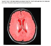
Rationale: To quantify the development of atrophy over time in super refractory status epilepticus (SRSE). Methods: We identified all episodes of SRSE treated between 2001 and 2013 at Mayo Clinic. Patients with 1) initial magnetic resonance imaging (MRI) study performed within 2 weeks of onset of SRSE, 2) repeat MRI within 6 months of the resolution of SRSE, and 3) a minimum of one week between scans were included. We measured the ventricle brain ratio (VBR) on T2/FLAIR images from disease onset and follow-up. Measurements were performed on axial FLAIR images with slice thickness of < 5 mm. The plane just superior to the caudate head was chosen as shown in the figure (Figure 1). Change in VBR (VBR delta) was calculated as a percentage of the starting measure. Days on anesthetic agents were recorded as a surrogate for duration of SRSE Results: Nineteen patients met inclusion criteria. Patients were 41 (25-68) years old and 52.6% were male. Anesthetic agents were required for 13 (5-37) days. Initial MRI was performed 2 (1-8) days from the onset of SRSE and the subsequent MRI was performed 11 (4-16) days from the resolution of SRSE with 40 (14-66) days between MRIs. The VBR delta ranged from -0.66% to -267% (median, -23.3%; lower quartile, -10.5%; upper quartile, -70.3%). There was a significant correlation between days on anesthetic agents and VBR delta (Spearman r=0.543; P=0.016) Conclusions: Development of atrophy occurred in nearly all patients with serial imaging in SRSE. Degree of atrophy appears to be related to duration of SRSE.
