ELECTROGRAPHIC NEONATAL SEIZURES AFTER NEWBORN HEART SURGERY
Abstract number :
A.04
Submission category :
Year :
2003
Submission ID :
4034
Source :
www.aesnet.org
Presentation date :
12/6/2003 12:00:00 AM
Published date :
Dec 1, 2003, 06:00 AM
Authors :
Uzma M. Sharif, Rebecca Ichord, J. William Gaynor, Thomas L. Spray, Susan Nicolson, Sarah Tabbutt, Gil Wernovsky, Robert R. Clancy Neurology, The Children[apos]s Hospital of Philadelphia, Philadelphia, PA; Cardiothoracic Surgery, The Children[apos]s Hospi
Neonatal seizures are an important early sign of acute encephalopathy and predictor of adverse neurodevelopmental outcome after newborn heart surgery. The contemporary occurrence of seizures is not known and their electrographic characteristics incompletely described. This study describes the contemporary characteristics of electrographic neonatal seizures (ENS) in a prospective cohort of infants with congenital heart disease (CHD) surgically repaired using cardiopulmonary bypass, with or without deep hypothermic circulatory arrest.
A cohort of consecutive infants undergoing newborn heart surgery was studied by continuous video-EEG monitoring for 48 hrs post-operatively. Records were reviewed for seizures every 24 hrs and ENS reported to clinical caretakers. Those with visually confirmed ENS were examined in detail to establish the time of first seizure, total number of ENS, site(s) of ENS origin and other characteristics.
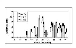
Over 180 infants underwent 48 hr. video-EEG monitoring from September 2001 to March 2003. ENS occurred in 21. None presented with clinically visualized seizures. A wide variety of different CHDs were represented. Affected infants were predominantly male (52%) and term (mean EGA 38 wks). The mean time to first ENS was 21 hrs (range 10-36). Mean total number of ENS over 48 hrs was 72 (range 1 to 217). The mean maximum number of ENS per hour was 7 (range 1 to 16). Post-op MRI was obtained in 17 of 21 cases and showed abnormalities in 15 of 17. The site of ENS origin was bilateral or multifocal in 10 infants with diffuse periventricular leukomalacia or global hypoxic-ischemic injury on MRI, while the origin of ENS correlated with site(s) of focal infarcts in 5 subjects. The figure shown is an example of the seizure profile in one subject with right temporal and left parietal infarcts. Clinically indicated EEG monitoring was continued beyond 48 hrs in 71% (15/21) and 6 infants continued to have ENS.
ENS, an important early marker of acute encephalopathy after newborn heart surgery, was common in a large, contemporary cohort of infants. The burden of ENS varied widely but many experienced numerous seizures. Seizures are a candidate outcome endpoint in future neuroprotection trials in this patient population.[figure1]
[Supported by: The Fannie E. Rippel Foundation and the American Heart Association]
