EVALUATION OF THE HYPOTHALAMI OF MENOPAUSAL WOMEN WITH EPILEPSY COMPARED TO MENOPAUSAL WOMEN WITHOUT EPILEPSY USING 1H-MRS
Abstract number :
1.215
Submission category :
Year :
2005
Submission ID :
5300
Source :
www.aesnet.org
Presentation date :
12/3/2005 12:00:00 AM
Published date :
Dec 2, 2005, 06:00 AM
Authors :
Blagovest G. Nikolov, Douglas R. Labar, and Cynthia L. Harden
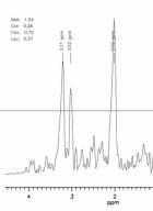
To determine if women with epilepsy and early onset menopause have abnormal neuronal integrity of the hypothalamus as measured by proton-magnetic resonance spectroscopy (1H-MRS). It is hypothesized that frequent interictal seizure disruption of the hypothalamus may lead to reproductive endocrine disorders in women. MRS may confirm hypothalamic abnormalities that are manifested through early onset menopause in women with epileptic seizures in comparison to age matched controls. 5 women (ages 52.62.7 years) presenting a history of epilepsy and early menopause were studied along with a set of matched controls. All data were acquired on a 3.0 Tesla Eclipse GE MRI scanner using a transmit/receive head coil. Metabolic ratios of NAA/CRE, NAA/CHO and CHO/CRE were calculated for the hypothalamus comparing values from the right side against the left as well as between the patient and control populations. In addition, ratios for the total hypothalamus were calculated and compared with normal grey matter within the same slice as shown in . No significant variations in metabolic ratios between patient and control populations were observed. Ratios of NAA/CRE, NAA/CHO, CHO/CRE for subject groups and regions of interest are listed below.
Controls Right Hypothalamus 1.870.20, 1.340.13, 1.400.15
Patients Right Hypothalamus 2.080.38, 1.530.28, 1.370.13
Controls Left Hypothalamus 1.850.25, 1.310.26, 1.430.21
Patients Left Hypothalamus 2.010.44, 1.480.43, 1.380.17
Controls Total Hypothalamus 1.860.21, 1.330.20, 1.420.17
Patients Total Hypothalamus 2.040.39, 1.500.34, 1.380.14
Controls Grey Matter 2.100.26, 1.540.27, 1.380.13
Patients Grey Matter 1.750.30, 1.540.21, 1.160.29 Spectroscopic studies have not focused specifically on the hypothalamus primarily due to the small structural size and resulting lack of signal to noise ratio. The resulting lack of significant MRS detectable metabolic variations between the patient and control populations may be due to the additional localization of reproductive dysfunction in the pituitary and peripheral glands in addition to the hypothalamus. Spectral quality at 3.0 Tesla in this small structure was more than adequate to provide accurate measures of metabolic ratios. Additional studies may provide more information on the location of metabolic abnormalities in patients with epilepsy that affect reproductive dysfunction.[figure1]
