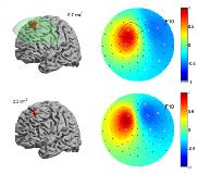
Extent of cortical sources visible on the scalp: effect of a subdural grid
Abstract number :
3.294
Submission category :
Late Breakers
Year :
2013
Submission ID :
1850018
Source :
www.aesnet.org
Presentation date :
12/7/2013 12:00:00 AM
Published date :
Dec 5, 2013, 06:00 AM
Authors :
N. von Ellenrieder, L. Beltrachini>, C. H. Muravchik, J. Gotman
Rationale: We analyzed the effect of a subdural electrocorticography (ECoG) grid on the scalp electric potential distribution, in order to assess the reliability of studies comparing simultaneous scalp and subdural recordings. In particular, we were interested in the validation of the minimum cortical extent of 10 cm2 reported to be necessary for observing epileptiform scalp EEG activity (Tao et al. Cortical substrates of scalp EEG epileptiform discharges. J Clin Neurophysiol. 2007; 24(2):96-100).Methods: Simulations were carried out in two detailed head models based on the Colin27 high resolution MRI segmentation of the Montreal Neurological Institute. More than 8 million tetrahedral elements were used (less than 1 mm side, with local refining near the cortex). Isotropic conductivity was assumed for the 8 tissues included in the model (skin and muscle, fat, bone, marrow, mayor blood vessels, CSF, gray matter, and white matter). The second model included the above model to which was added a non-conducting substrate of a subdural grid on the left frontal lobe (size 80x80 mm, 1.5 mm thickness, surrounded by at least 1 mm CSF in every direction), and four holes in the skull, near the corners of the grid (10 mm diameter). A cortical surface tessellated in more than 320,000 triangular elements was built following the mid surface of the cortex gray matter. Distributed sources of different extent (simulating epileptic spikes) were modeled, centered at 1000 points on the left hemisphere of this cortical surface. The electric potential generated on the scalp was computed with the Finite Element Method. Background brain activity noise was modeled with 6000 dipolar sources at random locations on the cortex.Results: Simulation results showed that the maximum amplitude of the scalp potential was attenuated for sources located under the non-conducting substrate, with attenuations of up to 5 or 6 times for sources located under the center of the grid. An increase in the scalp potential up to 3 or 4 times was observed for sources centered under the skull holes. Changes in the noise level on the scalp were much less pronounced, around 30%, also decreasing above the grid and increasing near the skull holes. Combining signal and noise results, we found that when the grid is not present, the scalp potential of cortical sources of 2 cm2 can attain the same signal to noise level as 10 cm2 sources located under the non-conducting grid. An example is shown in Figure 1. The variation of scalp potential amplitude seems to be important only for sources located precisely under the skull holes and the ECoG grid, thus the holes did not cancelled the effect of the grid. This also explains the small variation in noise level observed in the simulations. The background activity is generated by the whole brain, and only the small fraction generated near the grid and holes is affected by them.Conclusions: These results suggest that the reported cortical extent of 10 cm2 required to observe epileptiform discharges on the scalp EEG may be an overestimation, and deserves further study.
