FMRI OF DRUG-INDUCED AND SPONTANEOUS ABSENCE EPILEPSY IN AWAKE RATS
Abstract number :
1.115
Submission category :
Year :
2003
Submission ID :
3740
Source :
www.aesnet.org
Presentation date :
12/6/2003 12:00:00 AM
Published date :
Dec 1, 2003, 06:00 AM
Authors :
Jeffrey R. Tenney, Timothy Q. Duong, Jean A. King, Craig F. Ferris Center for Comparative Neuroimaging, Department of Psychiatry, University of Massachusetts Medical School, Worcester, MA
Typical absence seizures consist of multiple, brief impairments of consciousness with bilaterally synchronous 3 Hz spike and wave discharges (SWD) on electroencephalography (EEG). This study was undertaken to examine the feasibility of using functional magnetic resonance imaging (fMRI) to study both drug-induced and spontaneous SWDs in rodents. The [italic]a priori [/italic]hypothesis predicted an increase in BOLD signal in the corticothalamic circuit including the ventral posteromedial and posterolateral thalamus, reticular thalamic nucleus, and somatosensory cortex.
Rats were implanted with epidural EEG electrodes and secured in a MR compatible restrainer. [gamma]-butyrolactone (GBL) was used to induce absence seizures during blood-oxygenation-level-dependent (BOLD) fMRI, while WAG/Rij rats were imaged during spontaneous SWDs. EEG recording, with the animal in the magnet, was accomplished by using nonmagnetic epidural electrodes and non-conductive fiberoptic cables. Images were acquired with a 4.7T horizontal magnet. High resolution anatomical data sets were obtained using a fast spin echo sequence (TR=2.5s; TE=56ms; data matrix=256x256) and functional images with an echo planar imaging sequence (TR=1000ms; TE=25ms; matrix=128x128).
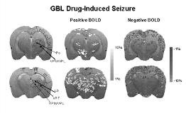
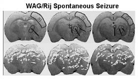
GBL caused robust changes in BOLD signal intensity in both cortical and thalamic structures. The thalamus had increased positive BOLD signal but there were few thalamic areas with negative BOLD signal. This is in contrast to cortical areas that showed a preponderance of negative BOLD signal during the SWDs. In addition, the sensory and parietal cortices had increased positive BOLD signal. Analysis of spontaneous SWDs in WAG/Rij rats showed similar positive BOLD signal changes in the cortex and thalamus, with asymmetric activation of several thalamic nuclei (right [gt] left). In addition, no negative BOLD signal occurred during spontaneous SWDs.
Using BOLD fMRI, we have demonstrated signal changes in brain areas of two frequently used animal models of absence epilepsy. The cortico-thalamic circuitry, critical for the formation of SWDs, showed robust BOLD signal changes during drug-induced and spontaneous seizures. These results corroborate previous findings from lesion and electrophysiological experiments and show the feasibility of non-invasively imaging absence seizures in fully conscious rodents. [figure1][figure2]
[Supported by: NIMH (R01-MH58700) and NINDS (F30-NS044672)]

