FOCAL MEG MONTAGES CAN DETECT FRONTAL LOBE SPIKES NOT SEEN IN ROUTINE EEG RECORDINGS
Abstract number :
2.223
Submission category :
Year :
2003
Submission ID :
1048
Source :
www.aesnet.org
Presentation date :
12/6/2003 12:00:00 AM
Published date :
Dec 1, 2003, 06:00 AM
Authors :
Jerry J. Shih, Deidre Devier, Elizabeth Bryniarski, Michael P. Weisend Department of Neurology, University of New Mexico School of Medicine, Albuquerque, NM; Department of Radiology, New Mexico VA Healthcare System, Albuquerque, NM
Patients with frontal lobe epilepsy (FLE) represent difficult diagnostic challenges. Interictal EEG performed in FLE often reveals no epileptiform abnormalities. Magnetoencephalography (MEG) is able to localize interictal spikes demonstrating agreement with invasive electrical recording and may be more sensitive than scalp EEG for the detection of epileptic discharges. In the present study, we compared MEG and EEG results from patients with FLE. We hypothesize that MEG is more sensitive than EEG in identifying interictal spikes in patients with FLE.
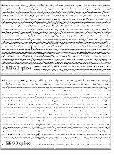
Simultaneously recorded MEG and EEG data were acquired from 12 patients diagnosed with FLE. The spontaneous data were displayed in three different configurations: EEG double banana montage (EOG, ECG, and 18 EEG channels), MEG double banana montage (EOG, ECG, and 18 MEG channels), and MEG focal montage (EOG, ECG, and 18 MEG channels from the identified frontal spike zone). An unblinded investigator reviewed the MEG and EEG records. This investigator identified segments of the records that contained spike(s). When a spike was identified on the record, a 10-second epoch containing the spike was printed for all three montages. The unblinded reviewer also identified segments that contained artifacts (i.e. eye blink, heart rate, movement, etc.) and printed 10-second epochs to act as foils. 477 10-second epochs (378 spike epochs, 99 foils), absent all identifying information, were provided to a blinded reviewer. The blinded reviewer was denied normal access to reference information in all epochs. The blinded reviewer recorded the presence of and/or number of spikes identified in each epoch. The results from this reviewer were tallied, returned to the unblinded investigators and matched with identifying information. Data were analyzed using the Wilcoxin Sign Rank Test to determine differences in detection of spikes from EEG, MEG focal montage, and MEG double banana montage.
The blinded reviewer detected significantly more spikes in the MEG focal montage compared to both EEG and the MEG double banana montage. The MEG double banana montage showed significantly less spikes than the EEG montage.
The use of focal MEG montages/derivations improves detection of spikes in FLE patients. Whether the improved detection rate is due to higher spatial clustering of sensors or to a higher signal-to-noise ratio remains to be elucidated.[figure1][table1]
[Supported by: NIH/NCRR 5P20RR15636 (JJS).]
