FOCAL REFRACTORY NONCONVULSIVE STATUS EPILEPTICUS AND ABNORMALITIES ON SEQUENTIAL MRI
Abstract number :
1.282
Submission category :
Year :
2004
Submission ID :
4310
Source :
www.aesnet.org
Presentation date :
12/2/2004 12:00:00 AM
Published date :
Dec 1, 2004, 06:00 AM
Authors :
Francisco Villalobos Chavez, and Juan Jesus Rodriguez Uranga
A series of transient alterations have been registered on MRI scanning studies of focal refractory nonconvulsive status epilepticus. We report a case and the abnormalities observed on sequential MRI. A 63-year-old female patient with a familial history of a sister and cousins suffering from epilepsy is admitted with confusional symptoms starting 72hrs before. Two similar episodes of 5 minutes each had occurred in the previous two years, which had recommended a cardiologic study, EEG and cranial MRI, all with normal results.
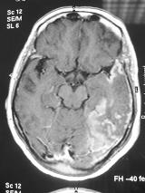
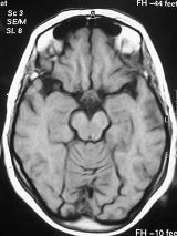
A new EEG was consistent with left temporal status epilepticus and the MRI scanning showed diffuse T2-weighted and FLAIR hyperintense images in the left temporal lobe without mass effect as well as gadollinium uptake areas in leptomeninges and cortex. CSF showed no cells and hyperproteinorrachia of 1 gr/dl. The symptoms continued for 10 days and the patient did not respond to antiepileptic drugs until the administration of steroids. A new MRI performed 10 days after resolution of symptoms showing a marked regression of the lesion following the administration of steroids. A new MRI performed after 3 months yielded normal results.[figure1][figure2] The case reported here supports the few previous reports on the occurrence of MRI abnormalities consistent with cytotoxic-vasogenic edema secondary to neuronal damage and rupture of the hematoencephalic barrier, respectively in relation with focal refractory nonconvulsive status epilepticus. Sequential MRI scanning, hyperproteinorrachia and the response to anti-edema therapy support our findings. Steroids may be a suitable therapeutic option in cases of refractory status epilepticus.

