GRAY MATTER DEFICITS CORRELATE WITH SEIZURE DURATION IN MESIAL TEMPORAL LOBE EPILEPSY WITH HIPPOCAMPAL SCLEROSIS
Abstract number :
2.298
Submission category :
Year :
2004
Submission ID :
787
Source :
www.aesnet.org
Presentation date :
12/2/2004 12:00:00 AM
Published date :
Dec 1, 2004, 06:00 AM
Authors :
1Jack J. Lin, 2Noriko Salamon, 1Rebecca A. Dutton, 1Agatha D. Lee, 1Jennifer A. Geaga, 1Kiralee M. Hayashi, 3Edythe D. London, 1Arthur W. Toga, 1Jerome Enge
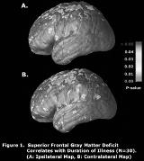
There is emerging evidence that patients with mesial temporal lobe epilepsy (MTLE) and hippocampal sclerosis (HS) have structural abnormalities that extend beyond the hippocampus. We report on a quantitative volumetric MRI study of patients with pathologically confirmed HS. Gray and white matter deficits were correlated with clinical variables. Quantitative volumetric analyses were performed on preoperative MRI brain scans of 15 left and 15 right MTLE (LMTLE/RMTLE) patients who underwent anteromesial temporal resection and have been seizure free for at least 2 years, and 20 age matched normal controls. MRI images were linearly registered to the International Consortium for Brain Mapping (ICBM) space. Tissue were classified into gray matter, white matter and cerebrospinal fluid. Lobar and whole hemisphere gray and white matter volumes were compared to normal controls. Regression analyses were performed to correlate tissue volumes with age, seizure duration, and history of febrile seizures. In LMTLE patients, a 35.6% average gray matter deficit was found in the left and a 34.6% average deficit in the right hemisphere (both P[lt].0001). In the RMTLE patients, a 39.4% gray matter deficit was found in the left and 40.4% in the right hemisphere (both P[lt].0001). There also was significant white matter loss in both MTLE groups with maximal deficit in the frontal and temporal lobes. Regression analysis showed that the age of the patient and duration of seizures were negatively correlated with gray matter volume ipsilateral (P[lt].04) and contralateral (P[lt].05) to the side of seizure onset. A history of febrile seizures did not correlate with tissue volumes. When data from the affected hemispheres of both MTLE groups were pooled, cortical gray matter deficits in superior frontal regions correlated significantly with seizure duration (P[lt].05, permutation test). Quantitative volumetric analysis of MTLE with HS showed widespread deficits in gray and white matter. Gray matter loss in superior frontal regions correlated with age and duration of seizures. This suggests MTLE may be a progressive disease involving multiple specific brain regions outside of the temporal lobe.[figure1] (Supported by Epilepsy Foundation and National Epifellows Foundation)
