Hippocampal Subfield Measurement and ILAE Hippocampal Sclerosis Subtype Classification With In Vivo 4.7 Tesla MRI
Abstract number :
1.247
Submission category :
5. Neuro Imaging / 5A. Structural Imaging
Year :
2018
Submission ID :
494463
Source :
www.aesnet.org
Presentation date :
12/1/2018 6:00:00 PM
Published date :
Nov 5, 2018, 18:00 PM
Authors :
Trevor A. Steve, University of Alberta; Ehsan Misaghi, University of Alberta; Tomasz A. Nowacki, University of Alberta; Laura M. Schmitt, University of Alberta; B. Matt Wheatley, University of Alberta; and Donald W. Gross, University of Alberta
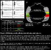
Rationale: Neuropathological studies have shown that hippocampal sclerosis (HS) consists of four distinct subtypes (ILAE Types 1-3 HS and No HS). However, HS subtypes currently can only be diagnosed by pathological analysis of hippocampal tissue resected during epilepsy surgery. In vivo diagnosis of HS subtypes holds potential to improve our understanding of these variants and enable prognostic information to be used for surgical decision-making. In the present study, we aimed to develop a method to characterize HS subtypes using in vivo MRI. Methods: Five subjects with TLE and unilateral HS (ILAE type 1 HS based on surgical pathology in all five cases) were compared with five healthy controls. We used a 4.7 T MRI system to acquire high resolution (0.26 x 0.34 x 1 mm3) MR Images of the hippocampus in each subject. In vivo-MRI diagnosis of ILAE HS subtypes was then determined in each patient by two methods: volumetric analysis and subfield area analysis along the hippocampal long axis (Figure 1). Results: Subfield volumetry demonstrated abnormalities in all five patients with three subjects demonstrating findings consistent with ILAE type 1 HS and two subjects with volumetry-defined atypical HS (one ILAE type 2 HS & one ILAE type 3 HS). Subfield area analyses demonstrated variability in ILAE HS subtype along the hippocampal long axis in several subjects (Figure 2). Conclusions: In the present study, hippocampal subfield volume abnormalities were observed for all subjects with agreement in ILAE subtype between histology and MRI observed in three out of five cases. The discrepancy of ILAE HS subtype observed for the remaining two subjects could be related to heterogeneity of HS subtypes along the long axis of the hippocampus. However, our results provide preliminary evidence that determining HS Subtype using high resolution in vivo MRI may allow preoperative diagnosis of ILAE HS subtypes. The heterogeneity of abnormalities observed with hippocampal subfield areas along the long axis of the hippocampus is consistent with previous autopsy studies and highlights the importance of studying the entire hippocampus in TLE patients. Funding: This work was supported by an operating grant held by DWG from the Canadian Institutes of Health Research (funding reference number 81083). TAS was supported by a post-graduate fellowship award from the Canadian League Against Epilepsy (CLAE). The funding sources had no involvement in this study aside from providing financial support.

.tmb-.png?Culture=en&sfvrsn=a09e81a3_0)