Histopathological Features of Hippocampal Sclerosis in Dual Pathology
Abstract number :
2.400
Submission category :
14. Neuropathology of Epilepsy
Year :
2018
Submission ID :
499556
Source :
www.aesnet.org
Presentation date :
12/2/2018 4:04:48 PM
Published date :
Nov 5, 2018, 18:00 PM
Authors :
Hajime Miyata, Research Institute for Brain and Blood Vessels; Saeko Sudo, Akita University School of Medicine; Yuichi Kubota, TMG Asaka Medical Center; Hidetoshi Nakamoto, TMG Asaka Medical Center; Ryoko Honda, National Nagasaki Kawatana Medical Center;
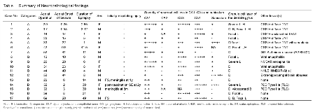
Rationale: Hippocampal sclerosis (HS) in patients with temporal lobe epilepsy (TLE) is characterized histologically by segmental neuronal cell loss with concomitant fibrillary gliosis in the hippocampal formation; however, its histogenesis remains elusive. HS occurs either isolated (i-HS) or in association with another epileptogenic principal lesion (dual pathology) (dp-HS), microscopically often showing indeterminate lesion patterns being difficult to classify within in the three ILAE types of HS. To elucidate the possible pathogenesis of HS, we performed comparative histological and immunohistochemical qualitative and quantitative evaluations of HS in surgically treated patients with TLE. Methods: Seventeen surgically resected specimens from patients with TLE were retrospectively chosen for this study from archival paraffin blocks. These include tissue in the following categories: (A) dp-HS with low-grade epilepsy-associated neuroepithelial tumor involving the hippocampus (n=7; age 2.9–52 years; mean 24.5±17.3); (B) dp-HS with encephalitis involving the hippocampus (n=4; age 16–44 years; mean 29.3±10.1); (C) dp-HS with extra-hippocampal brain abscess (n=1; age 52 years); (D) i-HS ILAE type 1 (n=5; age 20–69 years; mean 38.6±16.4 years). Sections of hippocampi were subjected to immunohistochemistry for NeuN, GFAP, chromogranin A, CD34 or CD3. Severity of neuronal loss in CA1–CA4 and subiculum was semi-quantitatively evaluated as following: (-) no obvious loss or gliosis only; (+) mild and/or focal; (++) moderate; (+++) severe (majority of neurons lost). Numbers of remaining neurons in CA1 per 1140 µm width and center of CA4 per 1 mm2 along with Voronoi diagram of remaining neurons were quantitatively evaluated. The mean numbers of remaining neurons and mean areas of Voronoi cells in each group were statistically compared by analysis of variance followed by a post hoc Turkey HSD or Games-Howell test for multiple comparisons (p<0.05). Results: In contrast to the stereotyped lesion pattern in i-HS ILAE type 1, lesion patterns in dp-HS did not fit into any ILAE type, showing incomplete, irregular, non-segmental neuronal loss in CA1 and/or CA4 in all but one case of extra-hippocampal brain abscess showing marked subicular involvement. More severe CA3 lesion than CA4 was observed in 1 tumor case. CA2 was relatively well preserved in all cases. Statistically significant difference was observed in mean number of CA1 neurons between i-HS group (14±9) and dp-HS with tumor group (90±61) (p=0.039) but not dp-HS with encephalitis group (36±28). No statistical difference was observed in mean areas of Voronoi cells between any groups. Conclusions: Pattern of hippocampal neuron loss in dp-HS is distinct from i-HS and can be classified as HS of indeterminate type, consistently characterized by incomplete or focal neuron loss in CA1 and CA4, particularly in cases with a second lesion diffusely involving the hippocampus. Nonetheless, there appears to be a selective vulnerability for tissue injuries, showing more severe neuronal loss in CA1 and/or CA4 followed by CA3, with relative sparing of CA2 and subiculum. Funding: None
