ICTAL SPECT-PET SUBTRACTION IN THE PRESURGICAL EPILEPSY EVALUATION
Abstract number :
1.121
Submission category :
Year :
2005
Submission ID :
5173
Source :
www.aesnet.org
Presentation date :
12/3/2005 12:00:00 AM
Published date :
Dec 2, 2005, 06:00 AM
Authors :
1Robert Knowlton, 1Jennifer Howell, 2Ojha Buddhiwardhan, 1Nita Limdi, 1R. Edward Faught, and 3Jorge Burneo
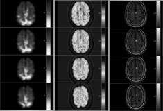
Subtraction techniques that use an interictal SPECT have improved sensitivity and specificity over ictal scans alone in localizing partial epilepsy for surgical treatment. In part this increase in sensitivity is related to increasing the magnitude of change due to epilepsy associated relative decreases in regional cerebral blood flow (rCBF) in the interictal state. Because metabolism is decreased more often and to a greater degree than rCBF, we investigated whether subtracting and interictal FDG-PET would increase the sensitivity for a localizing scan compared to ictal-interictal SPECT subtraction. Twenty-four patients who had ictal SPECT and FDG-PET scans as part of a larger prospective presurgical multimodality-imaging project were included in this preliminary observational study. Imaging techniques included:
1. High resolution MRI at either 1.5 or 3.0 T, using a standardized protocol optimized for extratemporal lobe epilepsy.
2. PET[ndash]5-10 mCi of FDG injected during the interictal state and scan performed on a high-resolution tomograph (FWHM=4-5 mm).
3. Ictal SPECT scans performed with HMPAO injections (within 30 s of seizure onset). Interictal injections were performed at least 24 hours before or after ictal injections.
4. Image registration, pixel intensity normalization, and image subtraction were performed in the Analyze[reg] Software environment on a Unix base workstation in identical fashion for both SPECT and PET subtraction. The mean age of patients was 28 years (range, 1-61 years; 9 females). Eleven patients had normal MRI (remaining had ambiguous, questionable, or nonlocalizing abnormalities). Epilepsy classification was: 15=extratemporal, 7=mesial-basal , and 2=lateral temporal. The sensitivity of detecting seizure associated rCBF increases for ictal-interictal SPECT subtraction was 8/21 (38%): 3=unifocal, 4=multifocal, and 1=lateralized. The sensitivity for ictal SPECT-PET subtraction was 20/24 (83%): 13=unifocal, 4=multifocal, 3=lateralized. Subtracting an interictal PET from an ictal SPECT scan may have a higher sensitivity for detecting a localizing rCBF than subtracting an interictal SPECT. Methodologically the technique novel and appealing, but further validation is warranted before consideration of clinical application.[figure1] (Supported by K23 NS02218, Robert Knowlton.)
