IMAGING ABSENCE SEIZURES USING fMRI
Abstract number :
C.07
Submission category :
Year :
2002
Submission ID :
112
Source :
www.aesnet.org
Presentation date :
12/7/2002 12:00:00 AM
Published date :
Dec 1, 2002, 06:00 AM
Authors :
Afraim Salek-Haddadi, Louis Lemieux, Martin Merschhemke, John S. Duncan, David R. Fish. Department of Clinical and Experimental Epilepsy, UCL Institute of Neurology, London, United Kingdom
RATIONALE: To identify and study the neural correlates of generalized spike-wave discharges (gSW) in man using simultaneous and continuous EEG-correlated fMRI.
METHODS: We studied a patient with intractable idiopathic generalised epilepsy using a 35-minute continuous whole-brain fMRI time-series during which 10-channels of scalp EEG were recorded simultaneously. This was enabled by an MR-compatible set-up with post-processing techniques to remove pulse (cardiobalistogram) (Allen PJ, Polizzi G et al., 1998) and imaging (Allen, Josephs, & Turner, 2000) artefact from the EEG. Four prolonged runs of 3Hz gSW (absence seizures) were fortuitously captured in their entirety. Fourier analysis was used to derive a running estimate of power spectral density at 3Hz which was convolved with a HRF to provide a regressor for statistical parametric mapping using the SPM99 Software.
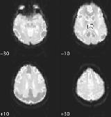
RESULTS: There were two distinct and highly significant patterns of BOLD change, time-locked to gSW. Activation was seen exclusively within the thalami bilaterally whilst profound deactivations were evident outside, symmetrically and over large areas of cortical grey matter with a midline frontal emphasis. All changes were consistent across seizures.
Activations (red-yellow) and deactivation (cyan-purple) are shown in the figure below as thresholded at the P[lt]0.05 level corrected for multiple comparisons, using separate colour-scales overlaid onto the structural EPI image.[figure1]
CONCLUSIONS: Despite an extensive body of work demonstrating thalamocortical mechanisms in animal models of gSW, surprisingly little has been established in man. PET and Doppler studies of cerebral blood flow and glucose metabolism during GSWD are conflicting, though reductions in both have been described(Theodore, Brooks et al., 1985;Nehlig, Vergnes et al., 1996). Our results provide direct evidence for a down-regulation of cortical activity during gSW, suggesting a key role for the thalamus. Moreover, the pattern of cortical involvement was in keeping with current density source reconstructions of gSW, suggesting frontal and occipital centres of gravity (Rodin, 1999).
References
Allen et al. Neuroimage 1998;8:229-239
Allen et al. Neuroimage 2000;12:230-239
Nehlig et al. 1996;16:147-155
Rodin et al. 1999;110:1868-1875
Theodore et al. 1999;35:684-690
[Supported by: Medical Research Council (UK).]
