IMPLICATIONS OF LOW NAA/CR IN HIPPOCAMPAL EPILEPSY
Abstract number :
G.04
Submission category :
Year :
2004
Submission ID :
5023
Source :
www.aesnet.org
Presentation date :
12/2/2004 12:00:00 AM
Published date :
Dec 1, 2004, 06:00 AM
Authors :
1Jullie W. Pan, 4Jung H. Kim, 2Aaron Cohen-Gadol, 2Dennis D. Spencer, and 3Hoby P. Hetherington
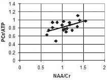
Many studies have reported declines in NAA/Cr in the hippocampus (ipsi-worse than contralateral) in MTLE. This measure is believed to reflect neuronal mitochondrial dysfunction. Although the sensitivity and specificity of the decline in NAA/Cr for seizure lateralization in MTLE is high ([gt]90%), few studies have reported how NAA/Cr relates to other measures, such as volumetry and neuropathology. Also, although methods exist to measure direct energetics (31P spectroscopic evaluations of PCr/ATP), no studies have evaluated both NAA/Cr and PCr/ATP in a common pool of MTLE patients. This abstract reports correlations of NAA/Cr with volumetry and neuropathology as well as 31P spectroscopic measures of energetics. We acquired high field structural imaging, 1H and 31P spectroscopic imaging studies in n=19 surgical MTLE patients together with neuropathologic measures. Hippocampal volumetry was performed using the methods of Watson and Jack. Neuropathology data included total gliosis, neuronal counts and quantitative glial fibrillary acidic protein (GFAP) staining. 1H spectroscopic imaging was performed using a 3D localized adiabatic moderate echo sequence. In a second study (acquired 30min after completing the 1H study), the 31P spectroscopic imaging was acquired. Analysis of the 1H and 31P data has been previously described. Pearson correlations were used for analysis, p[lt]0.05. A correlation between NAA/Cr with ipsilateral volumetry was significant (n=18) with r=0.50, p[lt]0.04. Neuropathologically, the NAA/Cr correlated with total GFAP (averaged across CA1-3, hilus) and total glial count (r=-0.54, p[lt]0.025 n=17 and r=-0.50, p[lt]0.03 n=19 respectively) while it did not correlate with neuronal counts. Finally, the hippocampal NAA/Cr significantly correlated (r=+0.52, p[lt]0.03, n=19) with amygdala PCr/ATP. The pes was similarly correlated. Declines in NAA/Cr are positively correlated with direct measures of energetic state, suggesting that if mitochonodrial functionality is low, then PCr/ATP is also low. The correlations with GFAP and total glial count suggest that NAA/Cr and energetics are more related to injury, rather than direct neuronal number. The positive correlation with hippocampal volumetry suggests that the remaining tissue in an atrophic hippocampus is energetically depressed in comparison to non-atrophic tissue. These multiple correlations between the energetics (PCr/ATP), volumetry, glial and GFAP counts with NAA/Cr suggest that neuronal loss and injury are linked with energetic depression.[figure1] (Supported by NINDS P01-39096 and R01-40550)
