INTERICTAL EEG-CORRELATED FUNCTIONAL MRI: A STUDY OF 50 PATIENTS WITH LOCALISATION-RELATED EPILEPSY
Abstract number :
1.245
Submission category :
Year :
2003
Submission ID :
4017
Source :
www.aesnet.org
Presentation date :
12/6/2003 12:00:00 AM
Published date :
Dec 1, 2003, 06:00 AM
Authors :
Afraim Salek-Haddadi, Beate Diehl, Martin Merschhemke, Khalid Hamandi, Louis Lemieux, David R. Fish Department of Clinical & Experimental Epilepsy, UCL Institute of Neurology, London, United Kingdom
EEG-correlated functional MRI allows for physiological imaging of spontaneous epileptiform activity, in vivo, across the entire brain, and with superior spatio-temporal resolution. Our aim was to characterise and map blood oxygen level dependent (BOLD) signal changes linked to interictal epileptiform discharges (IEDs), in a large group of patients with focal epilepsy.
50 patients with localisation-related epilepsy, independent of aetiology, and frequent IEDs on recent EEG were studied on a GE Horizon 1.5T scanner using whole-brain EPI (TE/TR 40/3000, 64x64 matrix). 700 scans were acquired continuously over 35-minutes. Ten channels of scalp EEG, plus ECG, were recorded simultaneously using an MR-compatible system, developed in-house, with on-line pulse and imaging artefact subtraction1.
IEDs were classified, labelled, and used to perform an event-related analysis of the fMRI data using SPM99. Images were realigned and smoothed. Event-related responses were modelled both flexibly and using a canonical heamodynamic response function (HRF). Results were thresholded for multiple comparisons.
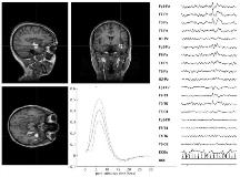
Good quality EEG was obtained in all patients and IEDs were successfully captured from 29. Of these, 12 (41%) had BOLD activations concordant with electroclinical data across a range of pathologies. 4 (14%) showed activation of uncertain significance and in 7 (24%) no activation was observed. In this group, there was a tendency to abnormal background rhythms, head motion, and subtle myoclonus. In 4 patients (14%) IEDs were too frequent for BOLD changes to be observed and in 2 patients, the study was terminated due to seizures. The HRF in patients with activation was in broad keeping with the [lsquo]canonical[rsquo] response, though some intersubject variability was evident. [figure1] EEG/fMRI in a 33 year-old female with left hippocampal sclerosis: BOLD activation, overlayed onto orthogonal slices (left), linked to runs of left temporal (F[sub]7[/sub]) spikes (see right). Fitted HRF is shown in lower middle.
Our experience indicates that simultaneous EEG/fMRI can provide novel localising information as to the irritative zone in selected patients, namely those with frequent stereotyped high-amplitude unifocal IEDs distinguishable from the background. Future studies need to address the pathophysiology of IEDs using more sophisticated models or consider alternative paradigms based on quantitative rather than qualitative EEG measures.
1. Allen PJ, Josephs O, Turner R. A Method for Removing Imaging Artifact from Continuous EEG Recorded during Functional MRI. Neuroimage 2000;12:230-9.
[Supported by: Medical Research Council (UK)]
