INTERICTAL GLUTAMATE LEVELS ARE DECREASED BUT THE RATE OF GLUTAMATE METABOLISM IN UNCHANGED IN TEMPORAL LOBE EPILEPSY AS REVEALED BY 1H MRS DURING INFUSION OF [U-13C] GLUCOSE
Abstract number :
2.378
Submission category :
Year :
2014
Submission ID :
1868930
Source :
www.aesnet.org
Presentation date :
12/6/2014 12:00:00 AM
Published date :
Dec 4, 2014, 06:00 AM
Authors :
Travis Losey, Brenda Bartnik, Daniel Ding, John Howe, Amul Shah, Tina Basak and Udochukwu Oyoyo
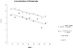
Rationale: Focal glucose hypometabolism in temporal lobe epilepsy (TLE) is associated with medical intractability, neurocognitive deficits and depression. The mechanisms by which hypometabolism affects metabolic processes are not well understood. Glutamate (glu) is the primary excitatory neurotransmitter in the brain and plays an important role in seizure generation and propagation. Glu is formed and regulated through several distinct but interrelated processes; oxidative metabolism of glucose to alpha-ketoglutarate and subsequently the glutamate-glutamine cycle, and anaplerosis within the astrocyte compartment. Extracellular glu concentrations have been found to be lower in the interictal state, with large amounts of glu released during focal seizures. However, increased glu has been seen in the epileptogenic hippocampus. Glu clearance after seizures is slowed in epileptic cortex and the amount of glutamate synthetase reduced in the involved hippocampus. Our study aimed to measure the rate of glu synthesis in the involved temporal lobe in the interictal state using a novel technique; the measurement of glu concentrations with 1H MRS during infusion of [U-13C] glucose, to better understand neuronal oxidative metabolism and the regulation of glu metabolism. Methods: Seven adults with unilateral TLE and 5 controls underwent structural MRI and 1H MRS using a 3.0T MR scanner. Single voxel spectroscopy (PRESS TR/TE = 2000/30 ms, number of averages = 160; voxel size 1.7 x 3.7x 1.7 cm) of the involved mesial temporal lobes were acquired at baseline and every 7.5 minutes during infusion of 20% w/v solution of 100% isotopically enriched [U-13C] glucose at 3mg/kg/min for 20 minutes. Subjects fasted for >12 hours prior to the study and were free of clinical seizures for > 24 hours before the study. LCmodel was used to obtain semi-quantitative metabolite levels and ratios. Custom software using Matlab and SPM 9 was used to obtain tissue composition and metabolic information from the same anatomical position. Subjects with epilepsy also underwent FDG PET. Statistical differences were determined using an independent samples Kruskal-Wallis test where p < 0.05 was considered significant. Results: Pre-infusion mean Glu/Cr ( 5.51 vs 4.05, p= 0.011) and glx/Cr (7.64/5.43 p=0.010) were higher in controls compared with TLE subjects. During infusion of [U-
