IS INTRAOPERATIVE ELECTROCORTICOGRAPHY RELIABLE IN CHILDREN WITH INTRACTABLE NEOCORTICAL EPILEPSY?
Abstract number :
A.02
Submission category :
Year :
2003
Submission ID :
4038
Source :
www.aesnet.org
Presentation date :
12/6/2003 12:00:00 AM
Published date :
Dec 1, 2003, 06:00 AM
Authors :
Eishi Asano, Krisztina Benedek, Aashit Shah, Csaba Juhasz, Otto Muzik, Diane C. Chugani, Sandeep Sood, Harry T. Chugani Pediatrics, Neurology, Radiology, Neurosurgery, Children[apos]s Hosp of MI, Wayne State Univ, Detroit, MI
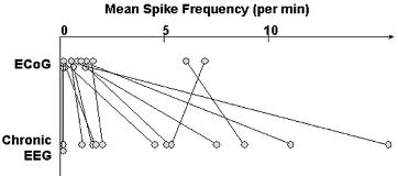
To study the relationship between spike frequencies during ECoG under general anesthesia and chronic subdural EEG recording in children with intractable neocortical epilepsy.
Thirteen children (age: 1.9-15.5 years) underwent a 10-minute intraoperative ECoG and chronic subdural EEG recording, using 64 to 120-channel subdural electrodes. During ECoG, isoflurane was maintained at 0.5-1.0% to minimize the effect on spiking. Using a spike detection program, the spike frequency during ECoG as well as during chronic subdural EEG was determined for each channel. The spike frequency for ECoG was compared to chronic subdural EEG for each patient (Wilcoxon signed-ranks test). The spatial pattern of spike frequency during ECoG was compared to chronic subdural EEG (Spearman[rsquo]s rank correlation). The relationship between the most frequently spiking electrode on ECoG and the ictal onset zone was analyzed in cases where at least three seizures were captured during chronic subdural EEG.
In 10/13 patients, the spike frequency was lower during ECoG than chronic subdural EEG (mean z = -6.1; p[lt]0.001). In one patient, there was no significant difference in the spike frequency between ECoG and chronic subdural EEG. In the other 2 patients, there were very few spikes during both ECoG and chronic subdural EEG, and appropriate comparison of spike frequency was not possible. A positive correlation was seen between the spike frequency patterns during ECoG and chronic subdural EEG (mean rho = 0.60; p[lt]0.001) in 9 cases, all of which exhibited frequent spikes ([gt]3 per minute) at least in one electrode during ECoG. In contrast, there was no correlation between the spike frequency pattern during ECoG and chronic subdural EEG (mean rho = 0.08) in the other 4 cases, all of which showed rare spikes ([lt]1 per minute) in all electrodes during ECoG. Ten patients had at least 3 seizures during chronic subdural EEG. All 6 children with frequent spikes ([gt]3 per minute) had the most frequently spiking electrode being a part of ictal onset, whereas all 4 patients with rare spikes ([lt]1 per minute) had the most frequently spiking electrode outside the ictal onset zone.
General anesthesia using isoflurane often decreases the spike frequency in children with neocortical epilepsy. Yet, the spike frequency pattern during ECoG is similar to that recorded during chronic subdural EEG, as long as frequent spikes ([gt]3 per minute) are seen in at least one electrode during ECoG. Rare spikes ([lt]1 per minute) in any electrode during ECoG could be considered as misleading background noise rather than indicators of the epileptic foci.[figure1]
