LARGER FDG-PET HYPOMETABOLISM ON MRI/PET FUSION STUDIES DETECTS THE EPILEPTIC FOCUS NON-INVASIVELY IN TUBEROUS SCLEROSIS COMPLEX PATIENTS
Abstract number :
2.460
Submission category :
Year :
2005
Submission ID :
5767
Source :
www.aesnet.org
Presentation date :
12/3/2005 12:00:00 AM
Published date :
Dec 2, 2005, 06:00 AM
Authors :
Sarat P. Chandra, Gary W. Mathern, Joyce Y. Wu, Susan Koh, Raman Sankar, and Noriko Salamon
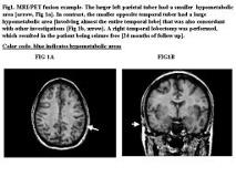
Surgery for intractable epilepsy in tuberous sclerosis complex (TSC) patients presents a challenge to localize the [apos][italic]epileptogenic[apos][/italic] tuber among the many without invasive EEG monitoring. This study evaluated the role of MRI/PET fusion in TSC patients to determine if this technique could potentially identify the epileptic tuber and focus. We reviewed 11 cases of TS operated for intractable epilepsy. In 5 cases MRI/PET fusion was utilized prospectively, and the other 6 were analyzed retrospectively. MRI/PET fusion images were processed at Vitrea workstation using Mirada fusion-7 software, and individual tubers were analyzed to discern if the area of FDG-PET hypometabolism was larger or the same size as the corresponding MRI tuber. None of the patients operated here required chronic EEG recordings, although all had scalp EEG telemetry and ECoG at surgery. In phase 1, we retrospectively reviewed 6 TSC cases to determine the utility of the MRI/PET fusion technique. We found that the resected epileptic focus and tuber were regions where PET hypometabolism was larger than the corresponding MRI tuber. Post-surgery seizure control occurred in all five patients where the enlarged PET area was removed. To verify this finding, we prospectively identified the epileptic tuber and focus in 5 other TSC cases. Analysis found again that the resected region was the area with expanded PET hypometabolism on MRI/PET fusion studies. Post-resection seizure control was obtained in 4 of the prospectively operated patients. MRI- PET fusion techniques were especially useful in identifying the epileptic focus and tuber in TSC patients when the FDG-PET hypometabolic area was larger than the anatomic MRI tuber compared with other [apos]non-epileptogenic tubers[apos]. While this results will need to validated in a larger surgical series, such an approach could reduce the need for chronic intracranial EEG recordings in TS patients undergoing epilepsy neurosurgery.[figure1] (Supported by NIH grant R01 NS38992.)
