Lobar Connectivity During Wakefulness and Sleep Studied Through Single Pulse Electrical Stimulation in Patients With Focal Epilepsy
Abstract number :
2.061
Submission category :
3. Neurophysiology / 3E. Brain Stimulation
Year :
2018
Submission ID :
501750
Source :
www.aesnet.org
Presentation date :
12/2/2018 4:04:48 PM
Published date :
Nov 5, 2018, 18:00 PM
Authors :
Anca Adriana Arbune, Emergency University Hospital, University of Medicine and Pharmacy “Carol Davila”, Bucharest, Romania; Ioana Mindruta, Emergency University Hospital, University of Medicine and Pharmacy “Carol Davila”, Buchares
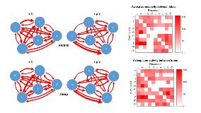
Rationale: Asymmetry in the human brain has been widely studied and is believed to be a determinant in the variation of individual functioning. Calculating the degree and direction of these variations could have important implications in the study of many neurological diseases. Because sleep is known as an activator of the epileptic brain, we aim to study the inter-lobar connectivity during wakefulness and sleep. Methods: We selected 17 consecutive patients with drug-resistant focal epilepsy who underwent presurgical evaluation with depth electrodes in 2016-2017 in our center. We implanted between 9 and 18 electrodes per patient, over a wide network, with at least 2 lobes explored. We performed single pulse electrical stimulation protocol (20 square, biphasic single pulses, pulse duration 0.3 ms and amplitude of 0.25 to 5 mA, in 0.25 mA steps) applied to adjacent contacts in eloquent cortex, during wakefulness and sleep. We recorded a total of 68288 responses out of which we selected and analyzed 20176 statistically significant responses (rho>0.4, p<0.05) from 84 structures. We used N-wavy ANOVA for further testing. Results: Looking at sleep-wakefulness inter-lobar intrahemispheric connections, we can observe that there are more statistically significant variations under the influence of sleep in the left hemisphere. Stimulation of the left temporal lobe during sleep determines greater responses in the left parietal (p<0.01) and insular cortex (p<0.01) as compared to wakefulness. On the other hand, the left fronto-parietal (p<0.05) and occipito-parietal (p<0.05) relationships during sleep reveal lower values in comparison with wakefulness state. The right hemisphere also exhibits some changes, sleep enhancing the occipito-insular connectivity (p<0.05) and diminishing the temporo-insular connectivity (p<0.05). Some of these intrahemispheric asymmetries have been previously described in resting-state fMRI in awake patients. Furthermore, we observed that the mean sleep-wake differences in the entire brain are 2.8 µV (p<0.001) when the epileptogenic zone is in the right hemisphere, and 6.2 µV (p<0.001) when the epileptogenic zone is in the left hemisphere. We also identified differences between lobes: when the epileptogenic zone is in the frontal lobe, the global mean sleep-wake difference is 6 µV (p<0.001), when in the temporal lobe the mean is 4 µV (p<0.001) and when in the insula the mean is 3.2 µV (p<0.001). Conclusions: We conclude that sleep has an influence on inter-lobar connectivity, especially in the left hemisphere, with possible implications in the evaluation of epileptic patients and other neurological afflictions. Global sleep-wake differences are also modulated by epileptogenic zone localization at lobar and hemispheric level. Additional data has to be collected in order to confirm these results. Funding: Romanian UEFISCDI research grants PN-III-P1-1.1-TE-2016-0706, PN-III-P4-ID-PCE-2016-0588, COFUND-FLAGERA II-SCALES and COFUND-FLAGERA II-CAUSALTOMICS
