Microstructural Imaging in Mesial Temporal Lobe Epilepsy: Role of Neurite Density and Myelination
Abstract number :
1.246
Submission category :
5. Neuro Imaging / 5A. Structural Imaging
Year :
2018
Submission ID :
493579
Source :
www.aesnet.org
Presentation date :
12/1/2018 6:00:00 PM
Published date :
Nov 5, 2018, 18:00 PM
Authors :
Gavin P. Winston, University College London; Sjoerd B. Vos, Centre for Medical Image Computing, University College London; Epilepsy Society, Chalfont St Peter; Benoit Caldairou, Neuroimaging of Epilepsy Laboratory, Montreal Neurological Institute and Hosp
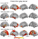
Rationale: In patients with mesial temporal lobe epilepsy (MTLE), neuroimaging studies demonstrate altered diffusion imaging parameters in ipsilateral temporolimbic and frontopolar cortices and widespread bilateral cortical thinning. Quantitative parameters that assume a single tissue type per voxel are sensitive but not specific to underlying microstructural changes. This study employed multi-compartment imaging techniques to explore the contribution of neurite density and myelination. Methods: 20 patients with MTLE (23-58 years, 11 male) and 20 matched controls (23-60 years, 11 male) were scanned. Quantitative parameters including neurite density, T1 and T2 relaxometry and myelin water fraction (MWF) were derived from NODDI and mcDESPOT models. Surfaces were extracted and parameters sampled at the midpoint between GM-CSF and GM-WM interfaces (intracortical) and 2mm below the GM-WM (subcortical). Linear models were used to assess groups differences, their relationship to epilepsy duration and the degree to which they explained conventional parameters. Results: Intracortical diffusivity was increased in ipsilateral temporolimbic and frontopolar cortices (Figure 1). Reduced neurite density was observed in mesial and basal temporal regions (PHG, fusiform). Diffusivity changes here and in the temporal pole and lateral temporal regions were explained by reduced neurite density. Increased T2 values compatible with gliosis explained the diffusivity changes in a similar distribution, whilst MWF was not significantly different.In subcortical white matter, fractional anisotropy showed a widespread reduction maximal in ipsilateral temporopolar and frontopolar regions (Figure 2). This was coincident with and partly explained by reduced neurite (axonal) density. These changes in diffusivity and neurite density were greater in patients with longer duration of epilepsy. An increase in radial diffusivity was detected in a similar distribution that was largely explained by the reduction in axonal density which in turn would be accompanied by loss of myelin. In support of this additional contribution by demyelination, a more confined reduction in MWF was seen in the ipsilateral temporal pole.Cortical thinning was seen bilaterally in temporolimbic and centroparietal cortices (Figure 1), but was largely independent of markers of neurite density and intracortical/subcortical myelination. Conclusions: Microstructural imaging suggests that neurite loss explains much of the intracortical diffusivity changes within the temporal lobe and that there is an additional contribution from demyelination in subcortical white matter. Greater changes in subcortical white matter were associated with longer disease duration. In contrast cortical thinning, particularly in more remote regions such as the centroparietal cortices, appears unrelated to neurite density or myelination. Histological confirmation of these findings is required. Funding: MRC (MR/M00841X/1), Epilepsy Society, UCLH/UCL NIHR Biomedical Research Centre

.tmb-.jpg?Culture=en&sfvrsn=2cd6b346_0)