NEURODEGENERATION DUE TO ELECTRODE IMPLANTATION IN FREELY BEHAVING ANIMALS
Abstract number :
3.059
Submission category :
Year :
2005
Submission ID :
5865
Source :
www.aesnet.org
Presentation date :
12/3/2005 12:00:00 AM
Published date :
Dec 2, 2005, 06:00 AM
Authors :
1,4Jonathan P. Mason, 1,4Nathalia Piexoto, 1,4Sridhar Sunderam, 2Ruchi S. Parekh, 1,3Steven L. Weinstein, 1,2Steven J. Schiff, and 1,4Bruce J. Gluckman
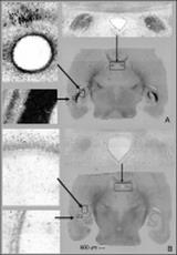
It is becoming increasingly clear from human studies that the implantation of depth electrodes can be associated with functional effects independent of electrical stimulation. Although such findings have been attributed to microlesions associated with electrode implantation, there is to our knowledge no prior work examining the anatomy of such microlesions. We present a neuropathological study of cell bodies and processes injured following depth electrode insertion into the hippocampus of rats. Two groups of male Sprague-Dawley rats (300g) were selected. The control group (n=6) was not implanted while the experimental group (n =7) was implanted bilaterally in the ventral hippocampus with electrodes constructed of stainless steel or steel coated with thin films of iridium oxide. These electrodes were placed so that they lay axially within the ventral hippocampus, within the curve of the CA3, by targeting a point near the tip of the enclosed blade of the dentate gyrus (Bregma referenced Paxinos and Watson 4th ed. atlas coordinates: AP = -5.15mm, ML = +/- 5.35mm, DV = -7.6mm). Implanted and non implanted animals were allowed free access to food and water for 10 days and then sacrificed and perfused with paraformaldehyde. All brains were sliced and stained with an Amino Cupric Silver stain to show neurodegeneration. We find significant neurodegeneration due solely to electrode implantation, without stimulation, for all electrode materials used (n = 4 animals). Electrodes inserted into the white matter of stratum radiatum caused local retrograde Wallerian-type degeneration of CA3 pyramidal neurons associated with transection of their axon collaterals and apical dendritic processes. Considerable degeneration of shaffer collateral and commissural fibers was also noted in the stratum pyramidale and oriens of CA1. Moreover, we found fiber degeneration in the alveus, fimbria and notably distally in the fornices (Figure 1). We present to our knowledge the first anatomical description of microlesion neuronal degeneration associated with the deep brain implantation of electrodes for use in epilepsy control. It is critical to distinguish functional effects of such lesions from the effects (functional or degenerative) from long term chronic stimulation of deep brain structures.[figure1] (Supported by NIH Grants R01EB001507, K02MH01493 and R01MH50006.)
