Presurgical Evaluation of Pediatric Epilepsy Patients: Relations Between EEG and MRI Localizations
Abstract number :
2.011
Submission category :
3. Neurophysiology / 3A. Video EEG Epilepsy-Monitoring
Year :
2018
Submission ID :
501474
Source :
www.aesnet.org
Presentation date :
12/2/2018 4:04:48 PM
Published date :
Nov 5, 2018, 18:00 PM
Authors :
Moritz Tacke, Dr. von Haunersches Kinderspital; Sophie Shen, Dr. von Haunersches Kinderspital; Jan Remi, Ludwig-Maximilians-University; Christian Vollmar, Ludwig-Maximilians-University; Soheyl Noachtar, Ludwig-Maximilians-University; Florian Heinen, Dr. v
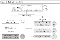
Rationale: Presurgical evaluation of pediatric patients aims to identify the epileptogenic lesion and its position relative to eloquent structures. The evaluation includes, among others, EEG video monitoring and MRI imaging. The results of both modalities are frequently incongruent, and the localizing value of EEG pathologies is uncertain. Methods: A total of 71 pediatric patients with MRI-positive lesions were included.The congruency between these lesions and the epileptic seizure patterns (ESPs) and interictal epileptiform discharges (IEDs) was analyzed. Results: Onset of ESP was over the lead corresponding to the MRI lesion in 77.5% of the seizures of patients with frontal lesions. The congruency rates for the other lesions were lower (temporal: 40.7%, parietal: 74.0%, occipital: 58.3%). The differences between the frontal and temporal ESP frequencies was statistically significant. A similar pattern emerged for the IED locations (frontal: 70.2%, temporal: 49.3%, parietal: 42.6%, occipital: 38.0%). Conclusions: Frontal lesions show the highest congruency between EEG video monitoring and imaging results. This is in contrast to the situation with adult patients, where temporal lesions show the highest congruency. Funding: None

.tmb-.png?Culture=en&sfvrsn=9ecabc0a_0)