Quantitative Spatiotemporal Characterization of Epileptic Spikes Using High-Density EEG: Differences Between NREM Sleep and REM Sleep
Abstract number :
1.134
Submission category :
3. Neurophysiology / 3A. Video EEG Epilepsy-Monitoring
Year :
2018
Submission ID :
496015
Source :
www.aesnet.org
Presentation date :
12/1/2018 6:00:00 PM
Published date :
Nov 5, 2018, 18:00 PM
Authors :
Xuan Kang, University of Wisconsin; Melanie Boly, University of Wisconsin; Cassie Williams, University of South Carolina; Graham Findlay, University of Wisconsin; Benjamin Jones, University of Wisconsin; Klevest Gjini, University of Wisconsin; Rama Magant
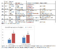
Rationale: Inter-ictal epileptiform discharges are useful markers to diagnose and localize epilepsy. Since the early days of clinical EEG, it was recognized that vigilance states have a profound effect on the frequency and distribution of inter-ictal epileptiform discharges. The advent of sophisticated methods such as high-density EEG and spatio-temporal statistics now allow for a systematic quantification of these differences. In this study, we look at differences in the distribution and duration of inter-ictal discharges during non rapid eye movement (NREM) sleep vs REM sleep in patients admitted to the epilepsy monitoring unit. Methods: Fifteen drug-refractory focal epilepsy patients were recruited from the Epilepsy Monitoring Unit (EMU) of the University of Wisconsin. 256 electrodes high-density EEG was applied in between the international 10-20 clinical electrode montage. 28 to 48 hour recordings were performed and the cleanest sleep epochs were selected for further analysis. Sleep was staged according to the American Academy of Sleep Medicine manual. NREM and REM sleep data were extracted, and board certified Epileptologists visually detected inter-ictal spikes. Among all the subjects, only six subjects displaying inter-ictal spikes during REM sleep were included in the analysis. EEG was averaged-referenced and epochs from -1000ms to 1000 ms after spike negative peaks were selected for analysis. Paired t-test were performed between spiking periods (data from 100ms before to 500s after spike peaks) and baseline (data from 1000ms to 300ms before spike peaks), and alpha level was set at 0.05, corrected for multiple comparison across 256 channels and across all time points. We quantified the average duration and number of channels significantly involved in spiking periods compared to baseline for each subject during NREM sleep and REM sleep. To detect group differences between NREM sleep and REM sleep we used a linear mixed effects model using sleep stage as a fixed effect and subject as a random effect. Testing for significance of models with either spike duration or number of channels involved was done with a likelihood ratio test with comparison to a null model. An additional permutation t-test was performed in each individual patient to assess the reproducibility of intra-subject differences in duration and number of channels involved in inter-ictal spikes during NREM sleep compared to REM sleep. Results: Patient demographics, including clinical EEG and imaging results, are displayed in Table 1. Subject P06 had only one spike during REM sleep. Channel results represent the number of channels significantly involved with each spike, and spike duration results represent the number of data points significantly involved with each spike. Figure 1 illustrates group-level t-test results, with a significantly higher number of channels involved in inter-ictal spikes and a significantly higher spike duration in NREM sleep compared to REM sleep. Overall effect of NREM versus REM sleep on duration of discharges was significant with a p-value of 5X10^-11, and for the number of channels it was 2x10^-5. Figure 2 and 3 also show consistent individual subjects results for significantly higher spike duration and significantly higher number of channels involved in spikes during NREM sleep compared to REM sleep. Conclusions: Our results suggest that epileptiform discharges occurring during REM sleep are shorter in duration and confined to a smaller scalp area compared to epileptiform discharges occurring during NREM sleep. Future studies will investigate the degree of spatial overlap between inter-ictal spikes present during NREM sleep versus REM sleep, and compared these measures with other markers of the epileptogenic zone. Funding: RO3 NINDS 1R03NS096379

.tmb-.png?Culture=en&sfvrsn=caf536a9_0)