SIMULATION STUDY SUGGESTS UNSAFE LEVELS OF SPECIFIC ABSORPTION RATE DURING SIMULTANEOUS EEG-MRI RECORDING
Abstract number :
2.229
Submission category :
Year :
2003
Submission ID :
3871
Source :
www.aesnet.org
Presentation date :
12/6/2003 12:00:00 AM
Published date :
Dec 1, 2003, 06:00 AM
Authors :
Leonardo Angelone, Andreas Potthast, Sunao Iwaki, Florent Segonne, Jack Belliveau, Giorgio Bonmassar Athinoula A. Martinos Center for Biomedial Imaging, Massachusetts General Hospital, Charlestown, MA
This study investigates the changes in Specific Absorption Rate (SAR) on human-head tissues during simultaneous Electroencephalography (EEG) and Magnetic Resonance Imaging (MRI) recording. Due to SAR considerations and risks of burns [1], this technology may present a safety hazard. We estimated the Radio Frequency power dissipated in the human head using a high-resolution head model generated from MRI data.
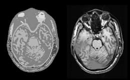
A High-Resolution head model (4395536 Yee cells) was realized applying a segmentation [2] to the anatomical MRI data of an adult male subject and selecting eight different types of tissue (Fig. left). The dimension of each cell was 1x1x1mm and the total volume considered, including the free space around the model, was 296*296*390mm. The surface coil used was a circular Perfect Electrical Conductor (PEC), ring-shaped and oriented in the coronal plane, with a diameter of 140mm and thickness of 1mm. The current source was placed on the lowest point of the ring and was a sinusoidal generator of 1A peak-to-peak amplitude with internal resistance of 50[Omega]. The birdcage coil was composed of 16 PEC rods (length 310mm), closed by two PEC loops at each end (diameter 260mm, thickness 1mm) and placed symmetrically around the head. A circular excitation was simulated driving each current generators placed on the center of each rod with a 1A peak-to-peak amplitude and a 22.5[deg] phase-shift between any two adjacent generators. All the simulations were conducted at the frequency of 300 MHz, (corresponding to a static MRI field of 7T), with and without EEG electrodes. The positions of the 124 electrodes and wires were digitized on the head of the subject and imported on the high-resolution model. The results were normalized to the region of interest (entire white matter with birdcage coil; posterior part of the white matter with the surface coil).
The results show that the ratio between whole-head SAR with EEG electrodes/leads and whole-head SAR without electrodes is equal to 1.26 with surface coil and 1.91 with birdcage coil. The figure (right) shows an axial MRI image with the position of SAR increase superimposed (red), with birdcage coil.
Our study shows that using non-magnetic metallic EEG electrodes and leads can increase the whole-head SAR to as much as two times with respect to the case without electrodes. Our results discourage the use of non-magnetic metallic EEG electrodes and leads for simultaneous EEG/MRI recording.
[italic][1] Chou, C.K., et al., Bioelectromagnetics, 1996. 17(3): p.195-208
[2] Dale, A.M., B. Fischl, and M.I. Sereno, Neuroimage, 1999. 9(2): p.179-94.[/italic][figure1]
[Supported by: Whitaker Foundation grant RG-99-0408.]
