SISCOM-GUIDED DEPTH ELECTRODES IN EPILEPSY SURGERY
Abstract number :
2.418
Submission category :
Year :
2004
Submission ID :
4867
Source :
www.aesnet.org
Presentation date :
12/2/2004 12:00:00 AM
Published date :
Dec 1, 2004, 06:00 AM
Authors :
1Jan Anders Ahnlide, 1Gunnar Skagerberg, 1Kristina Källén, 2Tove Hallböök, and 1Ingmar Rosén
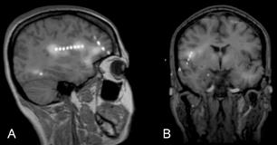
Subtraction ictal SPECT coregistered with MRI (SISCOM) has proved a valuable localizing method in epilepsy surgical workup. Our purpose is to demonstrate that SISCOM-guided stereotactic implantation of depth electrodes using a framed stereotactical system can localize the ictal onset zone in patients where SISCOM indicates areas difficult to assess with subdural electrodes. Implantation of depth electrodes with stereotactical technique, guided by SISCOM was performed in two patients with intractable epilepsy and normal findings on MRI. In both patients, SISCOM showed more than one area of hyperperfusion, of which at least one was unsuitable for investigation with subdural electrodes. Depth electrodes were implanted using SISCOM-guided stereotactical technique within the regions of hyperperfusion and also in the ipsilateral hippocampus. Multimodal fusion images were constructed, confirming the intended placement of the electrodes (fig). Invasive EEG recording localized the ictal onset zone in both patients. The first patient underwent a right sided temporal lobe resection. The second a right lateral temporal lobe resection under stereotactical surgical guidance. Neither patient is seizure free at short term post-operative follow up.[figure1] These cases suggest that combination of SISCOM, stereotactically implanted depth electrodes and stereotactically guided surgery can be reliably used in patients with intractable epilepsy where the suspected epileptogenic region is difficult to access with subdural EEG-electrodes or problematic to localize without guidance. Furthermore, in some cases this method might reduce the risk of complications by optimizing the electrode placement and thereby minimizing the number of intracranial electrodes required.
