SPATIAL DISTRIBUTION OF HIGH FREQUENCY ACTIVITY AND ITS RELATIONSHIP TO SURGICAL OUTCOME
Abstract number :
2.448
Submission category :
Year :
2005
Submission ID :
5755
Source :
www.aesnet.org
Presentation date :
12/3/2005 12:00:00 AM
Published date :
Dec 2, 2005, 06:00 AM
Focal high frequency activity (HFA) at seizure onset is associated with good surgical outcome in neocortical epilepsy but little information exists on widespread HFA and its relationship to ictal localization and surgical outcome. Subdural recordings (10 mm inter-electrode distance) from 4 patients with neocortical epilepsy were visually analyzed in bipolar and referential montages. Bandpass was set from 1.6 to 70/120/300 Hz (depending on sampling rate of 200/500/1000 Hz respectively) and 60 Hz notch filter was turned on. The range of implanted electrodes was 78-121. There were 19 seizures from the 4 consecutive patients with neocortical foci (2 frontal, 1 lateral temporal and 1 occipital). Patients #1 and #3 with frontal foci showed ictal HFA in [italic]all[/italic] the seizures (total 7 seizures, 3 from patient #1 and 4 from patient #3); none of the seizures from the other 2 patients had that finding. Only the 2 patients that had HFA were included in the subsequent analysis. Both were young females with normal MRI and PET, left frontal semiology, wide left hemispheric scalp ictal onsets, and inconclusive ictal SPECTs.
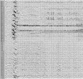
Initial HFA (iHFA) at seizure onset was in the range of 65-130 Hz (mean 89 Hz); it was distributed widely in 9-57 electrodes (median 45), representing 8-49% of implanted electrodes (median 37%), all within a contiguous cluster of 8-50 electrodes (median 32). On the contrary, the HFA was sustained (sHFA) focally in only 3-9 contiguous electrodes (median 5 electrodes) before evolving into lower frequency activity in the range of 20-80 Hz (mean 44 Hz). sHFA was seen in 2-8% of implanted electrodes (median 4%) with duration of 1-4 sec (mean 2.3 s). Figure shows widespread iHFA but focal sHFA in only 3 electrodes (PO39/40/47).
Patient #1 underwent left frontal resection inclusive of the entire sHFA area and 2 cm of adjacent iHFA area, had nonspecific gliosis on pathology, and has been seizure free for 14 months since surgery. Patient #3 underwent left fronto-parietal multiple subpial transactions only on the gyri that showed sHFA and has been seizure free for 3 months since surgery. When present in a given patient, the ictal HFA is consistent and occurs at the onset of each seizure, supporting the hypothesis that HFA is a unique electrical signature of the underlying epileptic brain. The widespread, non-localized distribution of iHFA is in contrast to previously published studies ([italic]J Clin Neurophysiol[/italic] 1992; 9:441-8, [italic]Brain[/italic] 2004; 127:1497-1506), probably due to the larger number of implanted electrodes in the current study. Inclusion of the sHFA areas in the surgical procedure appears to be critical in achieving favorable postoperative outcome.[figure1]
