Successful Surgery in a Patient With Bilateral Perisylvian Fast Activity at Ictal Onset
Abstract number :
1.369
Submission category :
9. Surgery / 9C. All Ages
Year :
2018
Submission ID :
502527
Source :
www.aesnet.org
Presentation date :
12/1/2018 6:00:00 PM
Published date :
Nov 5, 2018, 18:00 PM
Authors :
Ramya Raghupathi, Cleveland Clinic Foundation; Balu Krishnan, Cleveland Clinic; William Bingaman, Cleveland Clinic; Olesya Grinenko, Cleveland Clinic; Patrick Chauvel, Cleveland Clinic; and Juan Bulacio, Cleveland Clinic
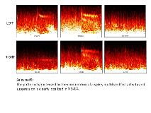
Rationale: Fast activity at ictal onset has been recognized as the main feature of epileptogenic zone from the beginning of the Stereo-EEG. Functional and anatomical connectivity of perisylvian cortical areas is known, however limited data is available showing bilateral perisylvian activation in focal seizures. We present a case in the setting of encephalomalacia lesion illustrating bilateral ictal onset in perisylvian cortex during invasive monitoring with good surgical outcome following unilateral resection. Methods: Not applicable Results: CASE DESCRIPTIONSixteen year old left-handed male with seizure onset at 6 years of age in the setting of left perinatal injury (no baseline deficits). Initial seizures were described as having an “a rush like feeling” in the head, followed by hyperventilation progressing to right hemi-body (arm and leg) numbness. This might evolve to right face clonic movement and further to involuntary laughter without loss of consciousness.Video EEG evaluation revealed no interictal epileptiform abnormalities and non-localizable seizures. MRI brain showed an extensive area of encephalomalacia involving left fronto-parietal operculum and dorsal left insular cortex. Ictal SPECT showed right frontal hyper-perfusion (injection at 20 secs) at OSH and left perisylvian hyper-perfusion at the Cleveland Clinic (9 secs). MEG at both institutions revealed tight clusters predominantly in the right insula-opercular regions and scattered dipoles in the left fronto-parietal-operculum. FMRI showed bilateral language areas activation and WADA right hemispheric language dominance.Given the discordant non-invasive data, intracerebral electrodes were implanted exploring bilateral perisylvian regions (left >right). Continuous slow background activity was noted in the inferior parietal lobule posterior to the encephalomalacia. Rhythmic sharp waves were commonly noted in the left fronto-opercular region. Ictal onset exhibited bilateral perisylvian low voltage fast activity (FA) followed by rhythmic high voltage sharp waves and slow activity on the left. His typical aura was elicited on stimulation of the left inferior parietal lobule (S' 7-8).The resection involved the left inferior Parietal lobule, extending anteriorly to left frontal-operculum. Postoperatively patient remains seizure (10mo). Time frequency analysis showed a combined pattern of a spike, multiband fast activity and suppression on the left (R’3’-4). Conclusions: The present of the FA is one of the main features of the epileptogenic zone, however it is not the only one. In our patient, the FA was bilateral, no difference in frequency range was noted visually but it was clearly demonstrated with time-frequency analysis. The resection on one side has achieved seizure control.The analysis of the FA should take into account the electrode placement (here not symmetrical), stimulation findings and the possibility that FA can be recorded outside of the epileptogenic zone. The frequency level of the FA cannot be considered in isolation. Analyzing the overall time-frequency pattern of the preictal/ictal transition is the way to define the epileptogenic zone.ReferencesBancaud J, Talairach J. La Stereo-electroence phalographie dans l’e´pilepsie: informations neurophysiopathologiques apporte es par l’investigation fonctionnelle stereotaxique. Paris: Masson; 1965.Bulacio J, Chauvel P, McGonigal A. Stereoelectroencephalography: interpretation. J Clin Neurophysiol. 2016;33:503-510.Grinenko O, Li J, Mosher J, Wang I, Bulacio J, Gonzalez-Martinez J, Nair D, Najm I, Leahy R, Chauvel P. A fingerprint of the epileptogenic zone in human epilepsies. Brain. 2018:141;117-131. Funding: Not applicable
