SURGICAL APPROACH IN PATIENTS WITH MESIAL TEMPORAL SCLEROSIS AND GROSS NEOCORTICAL SCARRING
Abstract number :
1.426
Submission category :
Year :
2003
Submission ID :
1918
Source :
www.aesnet.org
Presentation date :
12/6/2003 12:00:00 AM
Published date :
Dec 1, 2003, 06:00 AM
Authors :
Warren W. Boling, Adriana Palade, John Brick Department of Neurosurgery, West Virginia University, Morgantown, WV; Department of Neurology, West Virginia University, Morgantown, WV
To investigate the appropriate surgical approach in temporal lobe epileptics with mesial temporal sclerosis and prominent neocortical scarring.
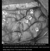
Four consecutive patients with prominent neocortical scarring and hippocampal sclerosis underwent surgery for intractable temporal lobe epilepsy at the West Virginia University Medical Center. Each patient had temporal lobe epilepsy defined by scalp EEG and seizure semiology. At surgery, extensive pial-dural adhesions over the temporal and frontal lobes were encountered upon reflection of the dura that was not evident on the preoperative MRI. Electrocorticography was recorded in each patient and a decision to perform a selective amygdalohippocampectomy or corticoamygdalohippocampectomy was based on results of the acute EEG recording. [figure1]
Despite extensive residual neocortical scarring, all the patients have had a marked reduction in seizure frequency or become seizure free with a selective amygdalohippocampectomy or anterior temporal corticoamygdalohippocampectomy.
Electrocorticography is a useful tool for formulating a resection strategy. Patients with extensive neocortical scar and temporal lobe epilepsy can realize an excellent result from surgery.
