The Clinical Utility of TMS in Localizing Language Specific Cortices: A MEG Comparison Study
Abstract number :
2.077
Submission category :
3. Neurophysiology / 3E. Brain Stimulation
Year :
2018
Submission ID :
502718
Source :
www.aesnet.org
Presentation date :
12/2/2018 4:04:48 PM
Published date :
Nov 5, 2018, 18:00 PM
Authors :
Kaylin Ryan, Rhodes College; Katherine Schiller, University of Tennessee Health Science Center; Roozbeh Rezaie, University of Tennessee Health Science Center, Memphis; Frederick Boop, University of Tennessee Health Science Center; James W. Wheless, Univer
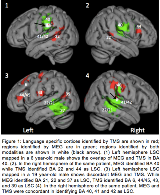
Rationale: Localization of eloquent cortices using non-invasive mapping methods may prove to be challenging in pediatric patients undergoing presurgical evaluation due to a lack of cooperation or ability. Increasingly, neuronavigated TMS has emerged as an effective, safe, and practical non-invasive mapping tool well suited for localizing language and motor cortices in children (Narayana 2015, J Child Neurol 30(9):1111-24). Previous work in pediatric patients has reported good concordance between magnetoencephalography (MEG) and TMS in assessing hemispheric dominance for language (Rezaie 2018, J Clin Neurophysiol, in press). Here we further investigated the compatibility of TMS and MEG language mapping by evaluating the spatial concordance of the two techniques in localizing language specific cortices (LSC). Methods: Patients with epilepsy or brain tumors (n=33, 5 to 36 years), identified through a retrospective review, underwent language mapping with TMS and MEG. Language mapping with MEG was performed using an auditory word recognition paradigm, andTMS language mapping was achieved using object naming task with pulses applied at a rate of 5 Hz TMS (80–100 V/m intensity) to the temporal and frontal cortices. TMS-elicited speech errors were categorized (speech arrest, semantic errors, and hesitations) and their cortical locations recorded. The concordance between MEG and TMS derived localization of LSC was examined by deriving statistical performance metrics for TMS including sensitivity, specificity, and diagnostic odds ratio (DOR). All MEG language activity sources and TMS speech error locations were overlaid onto patients’ anatomical MRI and transformed into MNI standard space for group analysis. Results: LSC identified by both TMS and MEG included Brodmann areas (BA) 21, 22, 39, 40, 41, and 42 in temporal and parietal lobes. To mitigate modality/task specific biases, LSC in frontal lobe (BA 6, 9, 13, 43-47) that were only identified by TMS were excluded. BA 22 and 21 were localized more frequently in MEG (73% and 53%) than in TMS (55% and 36%). BA 40 was activated more frequently in TMS (48%) than in MEG (23%). Thus, BA 22, 21, and 40 had the highest concordance of the 6 areas of interest, as seen through DOR values, presented in Table 1 with other statistical performance metrics. Conclusions: Temporal lobe LSC underlying linguistic processes were identified by both MEG and TMS and moderate concordance was noted between the two methods. Variability in spatial resolution and task differences may underlie divergences between the two techniques. However, the findings highlight how MEG and TMS language mapping can complement one another, particularly as it relates to localizing regions implicated in semantic processing. Studies focusing on optimization of testing parameters, including paradigm design, may further optimize TMS language mapping procedure and enhance its continued use as an addition to other non-invasive functional mapping techniques. Funding: Not applicable

.tmb-.png?Culture=en&sfvrsn=5ae4275f_0)