The Effect of Temporal Lobe Epilepsy on Memory Encoding Examined using Independent Component Analysis (ICA) and Granger Causality (GC)
Abstract number :
3.314
Submission category :
Late Breakers
Year :
2013
Submission ID :
1863581
Source :
www.aesnet.org
Presentation date :
12/7/2013 12:00:00 AM
Published date :
Dec 5, 2013, 06:00 AM
Authors :
A. M. Gregory, R. Nenert, J. Allendorfer, J. Szaflarski
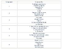
Rationale: Temporal lobe epilepsy (TLE) is frequently associated with memory impairments. Specifically, left TLE (LTLE) is commonly characterized by verbal and right TLE (RTLE) by visuospatial memory deficits. Standard general linear modeling (GLM) of scene encoding fMRI targeting visuospatial memory has shown bilateral activation modulated by side of seizure onset. The lateralization of the fMRI signals on standard GLM analyses are inconsistent with the results of memory testing but the reasons for this are unclear. ICA is a data-driven analysis method that is hypothesis-independent. It allows for identification of spatially and temporally independent components (ICs) that may overall contribute to the fMRI activation patterns observed using standard analysis methods. Thus, the goal of this study was to examine memory encoding using ICA and GC to identify the differential effects of L&RTLE. Methods: 55 epilepsy patients underwent fMRI using a scene-encoding task. 25 with LTLE and 19 with RTLE were kept for subsequent analyses. Patients were ages 19 to 66 (M = 38, SD = 12), 70% were male, with mean age of seizure onset at 20 years and a mean duration of epilepsy of 19 years. ICA identified 20 memory encoding components in each group. Some of the identified components were negatively related to the task (activated with control condition) these will not be discussed here. The results were subject to visual inspection and relevant components (positively correlated with the task) were retained for further examination using GC algorithm to identify casual influences between neural components with significance set at p<0.05, FDR corrected. Results: 3 ICs overlapped between L&RTLE these nodes are typically involved in memory processes (Table). 2 additional ICs were detected in LTLE patients and 1 in RTLE patients. Differences between L&RTLE have not been previously observed with standard GLM analyses. The 2 ICs in LTLE were observed in the L prefrontal cortices likely representing working memory and executive function contribution to task performance. RTLE patients showed activation in the occipital cortices, cuneus, fusiform and lingual gyri areas typically involved in visual imagery and verbal encoding. 4/5 ICs in LTLE and 4/4 ICs in RTLE showed significant causal influences with the origins in IC3 for both, L&RTLE. For LTLE, the casual influence is imposed on the L prefrontal/frontal cortices (unique ICs4&5) first with feedback to IC3 and feedforward to IC1 but none to the insular and superior temporal regions (IC4). In RTLE, casual influences are directed to each IC from IC3.Conclusions: L&RTLE affect memory encoding network differently. Consistent with previous studies, the organization of memory encoding is dependent on laterality of seizure focus and may be mediated by functional reorganization in chronic epilepsy. These findings may explain the differences in memory abilities between L&RTLE patients and highlight the modulating effects of epilepsy on the memory encoding network.
