THE RELATIONSHIP OF METABOLISM AND EPILEPTIC FOCUS IN TEMPORAL LOBE EPILEPSY
Abstract number :
2.428
Submission category :
Year :
2005
Submission ID :
5735
Source :
www.aesnet.org
Presentation date :
12/3/2005 12:00:00 AM
Published date :
Dec 2, 2005, 06:00 AM
Authors :
1,3Warren W. Boling, 2Adriana Palade, 1Michal Kraszpulski, and 3Gary Marano
FDG-PET has utility in lateralizing a seizure focus in temporal lobe epilepsy. However, the usefulness of FDG-PET to localize a seizure focus within a temporal lobe is not clear. The goal of this study is to determine the utility of the preoperative interictal FDG-PET to identify a mesial temporal epileptic focus, and guide a surgical approach. Consecutive subjects who underwent selective amygdalohippocampectomy (SAH) for temporal lobe epilepsy were analyzed. Exclusion criteria: prior resective surgery, follow-up under 1 year postoperative in seizure free subjects, and mass lesion. Inclusion criteria: FDG-PET performed as part of the preoperative evaluation, SAH surgery, and pre- and postoperative high resolution MRI to assess anatomic structures and the extent of resection.Two groups were compared. The free of seizures (FS) group represented a pure culture of subjects confirming the location of the seizure focus. A second group had continued seizures (CS) after surgery. Regions of interest (ROI) were defined on the MRI that were the mesial and lateral temporal structures. The FDG-PET was coregistered with the MRI and FDG activation was measured within each ROI. Normalized glucose metabolic rates for amygdala, hippocampus, and lateral cortex were calculated by dividing their average ROI activation values by that of the ipsilateral cerebellum.
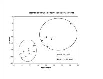
Postoperative seizure freedom defines successful removal of the seizure focus. Therefore, we hypothesize there will be a discernable difference in the metabolic activity of the mesial temporal structures in the SF versus the CS group. Eleven subjects were included in group FS. Six subjects were analyzed in group CS. In FS subjects, significant differences were found in the normalized FDG-PET metabolism for the mesial versus lateral temporal structures ipsilateral to the SAH resection (p [lt] 0.01). No significant difference was found in CS subjects (p=0.38). A high correlation was found between the normalized FDG-PET intensity in the mesial temporal structures and postoperative seizure freedom from SAH. (Figure 1). The results of this study demonstrate the utility of the preoperative interictal FDG-PET to identify a mesial temporal epileptic focus. The preoperative FDG-PET shows important differences in the metabolic activity of the mesial temporal structures in the seizure free versus the continuing seizure group. This finding has important implications in planning a surgical approach and better defining the localization of the seizure focus in temporal lobe epilepsy.[figure1]
