THE ROLE OF DIFFERENTIAL LOCAL TISSUE OXYGEN PRESSURE IN SPONTANEOUS[italic] IN VITRO[/italic] SEIZURES
Abstract number :
2.014
Submission category :
Year :
2005
Submission ID :
5318
Source :
www.aesnet.org
Presentation date :
12/3/2005 12:00:00 AM
Published date :
Dec 2, 2005, 06:00 AM
Authors :
1John R. Cressman, 1Jokubas Ziburkus, 1Kristen E. Johnson, 1,2Ernest Barreto, and 1,3Steven J. Schiff
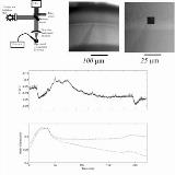
Recent simultaneous whole-cell measurements show that excitatory and inhibitory cells of the pyramidale and oriens layers of the CA1 exhibit interleaved patterns of activity during 4-AP induced seizures. Spontaneous seizures, as identified by a significant negative shift in extracellular voltage, occur after a period of high activity in the inhibitory cells. Seizures initiate as the inhibitory cells enter a transient state of depolarization block (DB) that is accompanied by a barrage of activity in the cells of the pyramidal layer. The origin of this layer specific blockade of cellular spiking activity is unknown, but it shares physiological similarities with hypoxic DB. Here we present a novel technique which permits the simultaneous measurement of electrical activity and quantitative oxygen measurement on the level of microdomains within neural tissue. We coat the tip of a microelectrode with a mixture of fluorescent dyes which permit the measurement of local tissue oxygen pressure along with extracellular or intracellular electrical potential. The dye cocktail is made by dissolving PVC, DOS - Bis, PtOEPKt - Pt(II) and BODIPY in the solvent tetrahydrofuran. Borosilicate glass micropipettes are dipped in the above cocktail, calibrated and inserted into tissue. Imaging is performed using a 580 nm excitation filter, a 590 nm dichroic mirror and the fluorescence light is filtered alternatively at 620 nm and 760 nm to image the BODIPY and PtoEPKt-Pt(II) respectively(see schematic). We simultaneously measured the local oxygen and extracellular potential in different strata in the CA1 region of the rat hippocampus during 4-AP induced seizures. Intracellular measurements reveal interleaving activity between excitatory cells in the st. pyramidale and inhibitory cells in the st. oriens. Simultaneous electrical and optical signals are displayed in figure 1 B and C respectively. Optical measurements display a decrease in the local oxygen pressure during the body of the seizure. We have invented a novel probe which is capable of simultaneously measuring extracellular potential and oxygen concentration at the same location. This will allow a detailed investigation of the role that oxygen plays on the level of microdomains of cortical layers or even near single cells in the formation and termination of seizures.[figure1] (Supported by NIH Grants: K02MH01493, K25MH01963, RO1MH50005.)
