THREE MRI PATTERNS OF CEREBELLAR ATROPHY IN PEOPLE WITH EPILEPSY
Abstract number :
1.213
Submission category :
Year :
2003
Submission ID :
1174
Source :
www.aesnet.org
Presentation date :
12/6/2003 12:00:00 AM
Published date :
Dec 1, 2003, 06:00 AM
Authors :
Michel J. Berg, Virginia P. Moreno, Lynn C. Liu Department of Neurology; Strong Epilepsy Center, University of Rochester, Rochester, NY
Cerebellar atrophy occurs to various degrees in people with epilepsy. Usually it is attributed to chronic medication use, although this mechanism has been debated. Coordination dysfunction has been correlated to cerebellar atrophy. Cerebellar atrophy has been postulated to worsen seizure control and severity as the cerebellum is generally thought to be an inhibitory structure.
We investigated 57 consecutive patients with available MRIs and video-EEG monitoring proven epilepsy. The degree of cerebellar atrophy was determined by making seven measurements on each of three coronal views (anterior, mid, and posterior) and two measurements on each of three sagittal views (mid left, midline, and mid right). The measurements were designed to define the widest distances between the folia and the degree of separation of the cerebellar margin from the dura. Values of zero were used as controls (i.e. no measurable distance between the folia or cerebellar margin should be present on the MRI.).
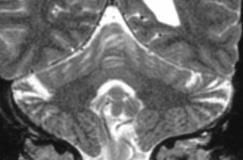
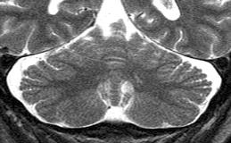
We observed three distinct MRI patterns of cerebellar atrophy in people with epilepsy: 1) folia atrophy (figure 1), 2) global atrophy without folia involvement (figure 2), and 3) a combination of both folia and global atrophy. Of the 57 patients, 10 (18%) had atrophy affecting only the folia, 2 (4%) had global atrophy alone and 8 (14%) had both folia and global atrophy. These different patterns of cerebellar atrophy would not have been apparent using total cerebellar volume measurements.
Three patterns of cerebellar atrophy occur in people with epilepsy: 1) folia atrophy, 2) global atrophy without individual folia involvement and 3) folia and global atrophy. Future analysis of the causes and consequences of cerebellar atrophy may be more revealing when segregated based on these patterns of atrophy.[figure1][figure2]

