UNILATERAL GIANT SOMATOSENSORY EVOKED FIELDS AS A LATERALIZING SIGN IN FRONTAL LOBE EPILEPSY
Abstract number :
1.001
Submission category :
Year :
2003
Submission ID :
2088
Source :
www.aesnet.org
Presentation date :
12/6/2003 12:00:00 AM
Published date :
Dec 1, 2003, 06:00 AM
Authors :
Valerie A. Carr, Susanne Knake, Hideaki Shiraishi, Deirdre M. Foxe, Keiko Hara, P. Ellen Grant, Donald L. Schomer, Blaise F. Bourgeois, Eric Halgren, Steven M. Stufflebeam Martinos Center for Biomedical Imaging, MGH/Harvard Medical School, Charlestown, MA
In patients (pts) with intractable focal epilepsies who are surgical candidates, MEG may be used to lateralize and localize the irritative zone and eloquent cortex. Somatosensory evoked fields (SEF) are often used as landmarks to localize the central sulcus and precentral gyrus. Here we report the use of SEF to lateralize the epileptogenic region in two pts with intractable frontal lobe epilepsy.
Pts were evaluated using 306-channel MEG and 64-channel EEG. Spontaneous data and median nerve SEF were obtained, and tibial nerve SEF were collected for one pt. Equivalent current dipoles (ECD) were calculated for interictal spikes (IIS) and SEF (N20m, P35m).
Case 1: 37 y old, rh female with two seizure types: (1) loss of vision [rarr] inability to speak and move, and (2) right shoulder twitching [rarr] loss of consciousness. Routine EEG, PET, SPECT and neurological examination were normal; EEG showed right temporal and bicentral spikes; MRI showed enlarged ventricles. ECD calculated for IIS localized to the right somatosensory cortex. Left median nerve stimulation showed a giant SEF response (218 fT/cm, 35ms), and was three times larger than that for the right median nerve (74 fT/cm, 35ms). ECD calculated from the giant SEF localized to the right central sulcus in the same vicinity as the IIS.
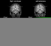
Case 2: 13 y old, rh female with simple motor seizures of unknown etiology and the following semiology: numbness right foot, hand, or head [rarr] clonic movements right arm and leg. EEG showed right centro-parietal and left para-sagittal spikes; MRI was normal. ECD of IIS clustered in the left hand and foot areas of the precentral gyrus. Right median nerve stimulation showed a giant SEF eight times larger in amplitude than that for the left median nerve (see figure). Right tibial nerve SEF (161 fT/cm, 42ms) was remarkably larger than that for the left (10 fT/cm, 40ms). Dipoles fitted to the giant right median and tibial nerve SEF localized to the left hand and foot areas, respectively.
Giant SEF may occur bilaterally in idiopathic generalized epilepsy due to general loss of inhibition and resulting hyperexcitability of the cortex. Here the pts[rsquo] giant SEF were seen on the hemisphere of the irritative zone, most likely due to increased excitability in this specific region. For pts with a suspected epileptic focus in the central region and for whom semiology, EEG monitoring, and imaging methods do not give clear localizing/lateralizing findings, large interhemispheric differences in SEF may help to lateralize the irritative zone.[figure1]
[Supported by: MIND Institute]
