USE OF STEREOELECTROENCEPHALOGRAPHY (SEEG) ELECTRODES TO PERFORM MULTIPLE RADIO-FREQUENCY THERMOLESIONS OF EPILEPTIC FOCI
Abstract number :
2.452
Submission category :
Year :
2003
Submission ID :
3713
Source :
www.aesnet.org
Presentation date :
12/6/2003 12:00:00 AM
Published date :
Dec 1, 2003, 06:00 AM
Authors :
Marc Guenot, Jean Isnard, Philippe Ryvlin, Catherine Fischer, Francois Mauguiere, Marc Sindou Dept of Functional Neurosurgery, Hop; P. Wertheimer, Lyon, France; Dept of Epileptology and Functional Neurology, Hop; P. Wertheimer, Lyon, France
Invasive recordings may be required in some cases of epilepsy before any surgical decision can be taken. We usually perform StereoElectroEncephaloGraphy (SEEG) recordings to achieve this goal (1). SEEG consists of stereotactic implantation of several depth electrodes in the brain, in order to identify exact location(s) of epileptogenic foci, as well as the propagation pathways of discharges. Electrodes (Dixi Medical, Besancon, France) are implanted in a stereotactic orthogonal way, using both MRI and angiography. Each electrode has from 5 to 15 contacts; each patient has 11 electrodes on average (from 5 to 16). The aim of this work is to demonstrate how it is possible to take advantage from these chronically implanted depth electrodes to perform RadioFrequency (RF) -thermolesions of epileptic foci and networks of the patients.
Ten adult epileptic patients (1 hippocampal sclerosis, 8 cortical dysplasias, 1 temporal neocortical focus) underwent multiple cortical RF-thermolesions made by means of chronically implanted SEEG electrodes between june 2001 and september 2002 at the department of functional neurosurgery of the university of Lyon. The lesions were always bipolar, they were done between two contiguous contacts of each selected eletrode. They were performed using a 50 volts, 110 mA current, applied during 10 to 30 sec (according to the delay of occurence of an abrupt decrease of the current intensity). 2 to 16 lesions were performed in each of the 10 patients, without any anesthesia, and under clinical and electrophysiological monitoring. An MRI examination was performed two months later in each case.
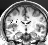
Each of these bipolar thermocoagulations proved to be able to create 4 to 7 mm diameter parenchymal lesions. [figure1]No general or neurological complication occured during the procedures, excepted a transient heat sensation of the head in one case. One transient post-procedure side-effect was reported (paresthetic sensations in the mouth). One patient (hippocampal sclerosis) was seizure free, six patients experienced a 80% improvement, three patients had little or no improvement of their seizures.
The technique of RF-thermolesions made by means of chronically implanted SEEG electrodes proved to be feasable, efficient, reliable, and safe. It appears to provide promising epileptological results, especially in term of palliative treatment of non removable epileptogenic foci.
(1) Guenot M, Isnard J, Ryvlin P, Fischer C, Ostrowsky K, Mauguiere F, Sindou M. Neurophysiological monitoring for epilepsy surgery: the Talairach SEEG method. Stereotact Funct Neurosurg 2002, 73:84-87.
