AUTOMATIC DELINEATION OF FOCAL CORTICAL DYSPLASIA LESIONS ON MRI
Abstract number :
1.074
Submission category :
Year :
2005
Submission ID :
5126
Source :
www.aesnet.org
Presentation date :
12/3/2005 12:00:00 AM
Published date :
Dec 2, 2005, 06:00 AM
Authors :
Olivier Colliot, Tommaso Mansi, Neda Bernasconi, Véronique Naessens, Dennis Klironomos, and Andrea Bernasconi
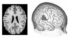
MRI has allowed the recognition of focal cortical dysplasia (FCD) in an increasing number of patients. The precise delineation of FCD is important since freedom from seizures after surgery is closely related to the extent of resection of the pathological tissue. FCD lesions are difficult to delineate on MRI due to their ill-defined boundaries and the complexity of the cortex. We previously developed an automatic classifier that successfully detected FCD in the majority of cases (Antel et al. [italic]Neuroimage[/italic] 2003; 19: 1748-59). However, the classifier sampled on average only 20% of the lesion extent. Our purpose was to design and evaluate an automatic method for accurate delineation of FCD on MRI. We studied 18 patients with MRI-visible FCD and partial epilepsy. High-resolution MRI was acquired on a 1.5T scanner using a T1-FFE sequence (1mm[sup3] isotropic voxels). To automatically delineate the FCD, we used a 3D deformable model that makes a surface evolve from an initial position towards the boundaries of the lesion. The initial position was obtained using our previously developed classifier under supervision of an expert observer. The deformable model evolution was driven by feature maps representing three known visual characteristics of FCD (cortical thickening, blurred GM/WM transition, and hyperintense signal), in order to distinguish the lesion from normal tissues. Figure 1 presents an example of automatic delineation. Lesions were manually delineated independently on the MRI by two trained raters to validate the automatic segmentation. Agreement between raters and between automated and manual delineations was assessed using Dice[apos]s similarity index. The mean interrater similarity was 0.62[plusmn]0.19, indicating a substantial agreement. The similarities between automated delineation and the manual labeling of each of the two raters were 0.62[plusmn]0.16 and 0.63[plusmn]0.12 respectively, indicating also a substantial agreement. We demonstrated for the first time the ability of advanced segmentation techniques based on deformable models to automatically delineate FCD lesions on MRI. By being objective and reproducible, our approach overcomes the limits of manual tracing. Moreover, it has the potential to unveil lesional areas that could be overlooked by visual inspection. This new method may become a useful tool for surgical planning in epilepsy.[figure1] (Supported by Epilepsy Canada, Canadian Institutes of Health Research (#203707), Scottish Rite Charitable Foundation.)
